Vesalius and the Fabrica of 1543
In the summer of 1543 the Basel printer Johannes Oporinus issued a folio volume of more than six hundred pages under the title De humani corporis fabrica libri septem, “Seven Books on the Fabric of the Human Body.” Its author, Andreas Vesalius, was twenty-eight years old and held the chair of surgery and anatomy at the University of Padua. The book was dedicated to the Emperor Charles V, printed in a large and elegant roman type, and illustrated with more than two hundred woodcuts of a quality that no medical book had approached before. Its pages showed skeletons leaning on spades and contemplating skulls, flayed figures striding through an Italian landscape, and dozens of smaller cuts of bones, vessels, nerves and organs set into the text beside the passages that described them. What the woodcuts depicted, and what the text argued, was that the anatomy taught in the universities of Europe for more than a thousand years had been drawn largely from animals, and that the only reliable guide to the structure of the human body was the human body itself, opened and examined by the physician’s own hands. This article follows Vesalius from his schooling in Brussels, Louvain and Paris to the lecture theatre at Padua, describes how the Fabrica was made and what it contained, and traces the arguments it provoked and the line of anatomical work that led from it to the discovery of the circulation of the blood.
Anatomy Before Vesalius
The anatomy that Vesalius inherited rested on the writings of Galen of Pergamon, the Greek physician who served the gladiators of his native city and later the imperial household in Rome in the second century AD. Galen was an exceptionally skilled and prolific investigator. He demonstrated that the arteries carry blood rather than air, showed by cutting the recurrent laryngeal nerve of a squealing pig that the voice depends on the nerves running to the larynx, and described the bones, muscles and nerves with a precision that later readers found hard to fault. His method, however, was constrained by Roman custom, which forbade the dissection of human bodies. Galen therefore dissected animals: pigs, dogs, oxen and, above all, the Barbary macaque, whose anatomy he believed to be closest to that of man. He supplemented these dissections with what he could observe in wounds, in the exposed bones of unburied corpses, and in the famous occasion, recorded in his own writings, when he studied a skeleton washed out of a grave by a flooding river. Where the anatomy of the ape or the ox differed from that of a human being, Galen’s descriptions carried the animal’s structure into the medical literature as though it were human.
Galen’s works passed through Byzantine, Syriac and Arabic hands before returning to Latin Europe, first in medieval translations from Arabic and then, from the late fifteenth century onward, in new Latin versions made directly from the Greek by humanist scholars. By the early sixteenth century his authority in the medical faculties was very nearly absolute. When human dissection was revived in the Italian universities in the late Middle Ages, it served mainly to illustrate the texts rather than to test them. The standard manual for some two centuries was the Anathomia of Mondino de’ Luzzi of Bologna, written in 1316, a short guide arranged in the order in which a body decayed, beginning with the abdomen, then the chest, then the head and the limbs. In the customary public dissection the professor sat in a raised chair reading aloud from Galen or Mondino, while a barber-surgeon did the cutting below and a demonstrator pointed with a rod to the parts as they were named. A woodcut in the 1493 Italian edition of the Fasciculus medicinae shows exactly this arrangement, with the learned man elevated above the corpse and the menial work left to others. Under such conditions discrepancies between book and body were easily explained away, if they were noticed at all, as defects in the particular cadaver or as changes in the human frame since antiquity.
There were signs of change before Vesalius. Jacopo Berengario da Carpi at Bologna published commentaries on Mondino in 1521 and 1522 that included his own observations and some crude but genuinely observational illustrations. Leonardo da Vinci had made hundreds of anatomical drawings of extraordinary penetration in the years around 1510, though they remained in private hands and had no influence on the teaching of anatomy. In Paris, the humanist physicians who edited and translated Galen were eager to recover the full extent of his anatomical work, including his treatise On Anatomical Procedures, which had been largely unknown in the medieval West. Their aim was to restore ancient knowledge, not to overturn it, but by drawing attention to Galen’s own insistence on dissection they created the conditions in which his conclusions could be checked.
Brussels, Louvain and Paris
Andries van Wesel, who latinised his name as Andreas Vesalius, was born in Brussels on 31 December 1514. His family came from Wesel in the duchy of Cleves and had served the house of Habsburg as physicians and apothecaries for several generations. His father, also named Andries, was apothecary to the Emperor Maximilian I and then to Charles V, and was often away with the court. The family house stood near the Gallows Hill outside Brussels, and Vesalius later recalled that the bodies of executed criminals left there for the birds were among the first human remains he saw. He was educated at the University of Louvain, where he studied the arts course, Latin and Greek, and in 1533 he went to Paris to study medicine.
In Paris Vesalius studied under Johann Guinter of Andernach, the translator of Galen, and Jacobus Sylvius, the most celebrated teacher of anatomy in the city and a devoted Galenist. Both men taught anatomy largely from the text. Vesalius found the formal dissections inadequate; the few that were held were brief and conducted by barbers, and he complained afterwards that apart from eight muscles of the abdomen, badly mangled, nobody had ever shown him a single muscle or bone, still less the course of the nerves, veins and arteries. He made up for this by his own efforts, collecting bones from the Cemetery of the Innocents and from the gibbet at Montfaucon, where he and his fellow students braved the dogs to study the skeletons of the hanged. He became so familiar with the bones that he could, he said, identify any of them blindfolded by touch. Guinter, in the 1536 edition of his Institutiones anatomicae, praised his young assistant as a man of great promise and exceptional knowledge of medicine.
The war between Charles V and Francis I of France forced Vesalius, as a subject of the emperor, to leave Paris in 1536. He returned to Louvain, where he and the physician Gemma Frisius took down a nearly complete skeleton from a gibbet outside the city, smuggled the bones back piece by piece, and assembled them at home under the pretence that he had brought the skeleton from Paris. He conducted a public dissection at Louvain, the first held there for eighteen years, and in February 1537 published his bachelor’s thesis, a paraphrase of the ninth book of the Almansor of the Persian physician Rhazes. Later that year he left for Italy.
The Chair at Padua
Vesalius arrived in Padua, the university of the Republic of Venice, in the autumn of 1537. He was examined for the doctorate on 5 December 1537 and passed with the highest distinction; on the following day the Venetian Senate appointed him explicator chirurgiae, lecturer in surgery, a post that carried with it responsibility for teaching anatomy. Padua offered advantages that Paris could not. The Venetian authorities were generous with bodies from the gallows, the faculty was large and international, and the university had long encouraged the practical side of medicine. Vesalius made use of all of this. He did his own dissecting, descending from the professor’s chair to the table, and he taught with the body open before the students rather than with a book in his hand.
He also taught with pictures. In 1538, to help his students follow the lectures, he published the Tabulae anatomicae sex, six large broadsheets printed in Venice. Three of them, showing the skeleton from the front, side and back, were drawn by Jan Steven van Calcar, a Netherlandish pupil of Titian who was working in Venice; the other three, showing the veins, arteries and organs, were drawn by Vesalius himself. The Tabulae were still largely Galenic in content. They showed, for example, a five-lobed liver, as in the dog or the ape, and the rete mirabile, a network of blood vessels at the base of the brain that Galen had described and that exists in oxen and sheep but not in humans. They were widely copied and pirated across Europe within a few years, which showed both how great the demand for anatomical images was and how little control an author had over his work once it left the press.
In the same year Vesalius issued a revised edition of Guinter’s Institutiones, and in 1539 a letter on venesection, the Epistola docens venam axillarem dextri cubiti in dolore laterali secandam, which intervened in a heated contemporary dispute over the site from which a patient should be bled in pleurisy. To settle the question he followed the veins in the body, and in doing so began to observe the valves and branches that the texts described imperfectly. Around this time a Paduan judge, Marcantonio Contarini, began to deliver the bodies of executed criminals to him, sometimes delaying the execution to suit the timetable of his teaching, which gave Vesalius a supply of fresh material far greater than any anatomist had previously enjoyed.
The decisive turn came gradually between 1538 and 1541. As Vesalius dissected more human bodies and compared them, sometimes side by side, with the bodies of apes and dogs, he found that many of Galen’s statements held true only of the animals. In January 1540 he gave a celebrated course of dissections at Bologna, at the invitation of the students there, which was recorded in detail by a German student, Baldasar Heseler, whose notes survive. Heseler described how Vesalius set the skeleton of a man beside that of a monkey and showed the audience the differences, and how he argued openly with the Galenist professor Matteo Corti, who lectured from the text while Vesalius demonstrated on the body. About the same time Vesalius was working on a new edition of Galen’s anatomical works for the Giunta press in Venice, a task that obliged him to read Galen line by line and to set each claim against what he had seen. By 1541 he had concluded that Galen had never dissected a human body and that his anatomy, for all its brilliance, was in large part the anatomy of the ape.
Making the Fabrica
The book in which Vesalius set out his conclusions was prepared in Padua and Venice between about 1539 and 1542. The drawings were made by artists working in Venice, almost certainly from dissections that Vesalius performed and supervised. The identity of the principal artist has been debated for centuries. Calcar is the traditional candidate, and the sixteenth-century art historian Giorgio Vasari attributed the drawings to him; others have proposed artists from the circle of Titian, or a group of draughtsmen working under Vesalius’s direction. No single attribution has commanded agreement, and the style varies enough across the book to suggest more than one hand. What is clear is that Vesalius regarded the illustrations as integral to the argument, specified what each should show, and labelled them with letters keyed to the text.
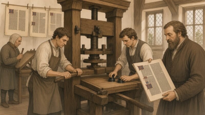
The drawings were transferred to pearwood blocks and cut in Venice by woodcutters of great skill. The blocks, together with Vesalius’s manuscript, were then carried over the Alps to Basel by the Milanese merchants of the Danoni firm, with a covering letter from Vesalius to Oporinus dated 24 August 1542 that survives in print at the front of the book. In it he asked the printer to follow the layout precisely, to set the letters in the figures with care, and to make sure that the impressions were clear. Oporinus, who had been professor of Greek in Basel and secretary to Paracelsus, was a printer of learned books with a reputation for accuracy. Why Vesalius chose a press so far from Padua is not certain, but Basel was a great centre of humanist printing, Oporinus was a meticulous craftsman, and a printer outside Venetian territory may have offered better protection against piracy. Vesalius himself travelled to Basel in early 1543 to oversee the printing, and he stayed long enough to dissect the body of an executed criminal, Jakob Karrer von Gebweiler, whose skeleton he assembled and presented to the university, where it is still preserved.
The Fabrica appeared in June 1543, the date given in its colophon. It is a large folio, about forty-three centimetres tall, of some 660 pages. At almost the same time Oporinus printed the Epitome, a much shorter companion in very large format intended for students and for those who could not afford the full work. It had a sequence of full-page figures and only a few pages of text, and its plates were designed to be cut out and assembled into a layered paper model of the body. The Fabrica was dedicated to Charles V; the Epitome to his son, the future Philip II of Spain. Vesalius presented a specially coloured copy of the Fabrica to the emperor, and another illuminated copy of the Epitome on vellum survives.
The Frontispiece and the Portrait
The title page of the Fabrica is one of the most famous images in the history of science. It shows a crowded anatomy theatre, built of timber in a classical architectural setting, packed with spectators who lean over the rails and push forward to see. At the centre Vesalius himself stands at a table on which lies the body of a woman, her abdomen opened. He has one hand inside the body and the other raised, as though addressing the audience. Above him a skeleton holds a staff, and the professor’s chair, which in older images dominated the scene, has disappeared; the barbers who once did the cutting sit beneath the table, sharpening razors, as though their role had been taken over. A dog and a monkey appear in the crowd, reminders of the animal dissections against which the book was directed. The scene is not a record of a particular occasion but a manifesto: anatomy was to be done by the anatomist, on the human body, in public, before witnesses who could see for themselves.
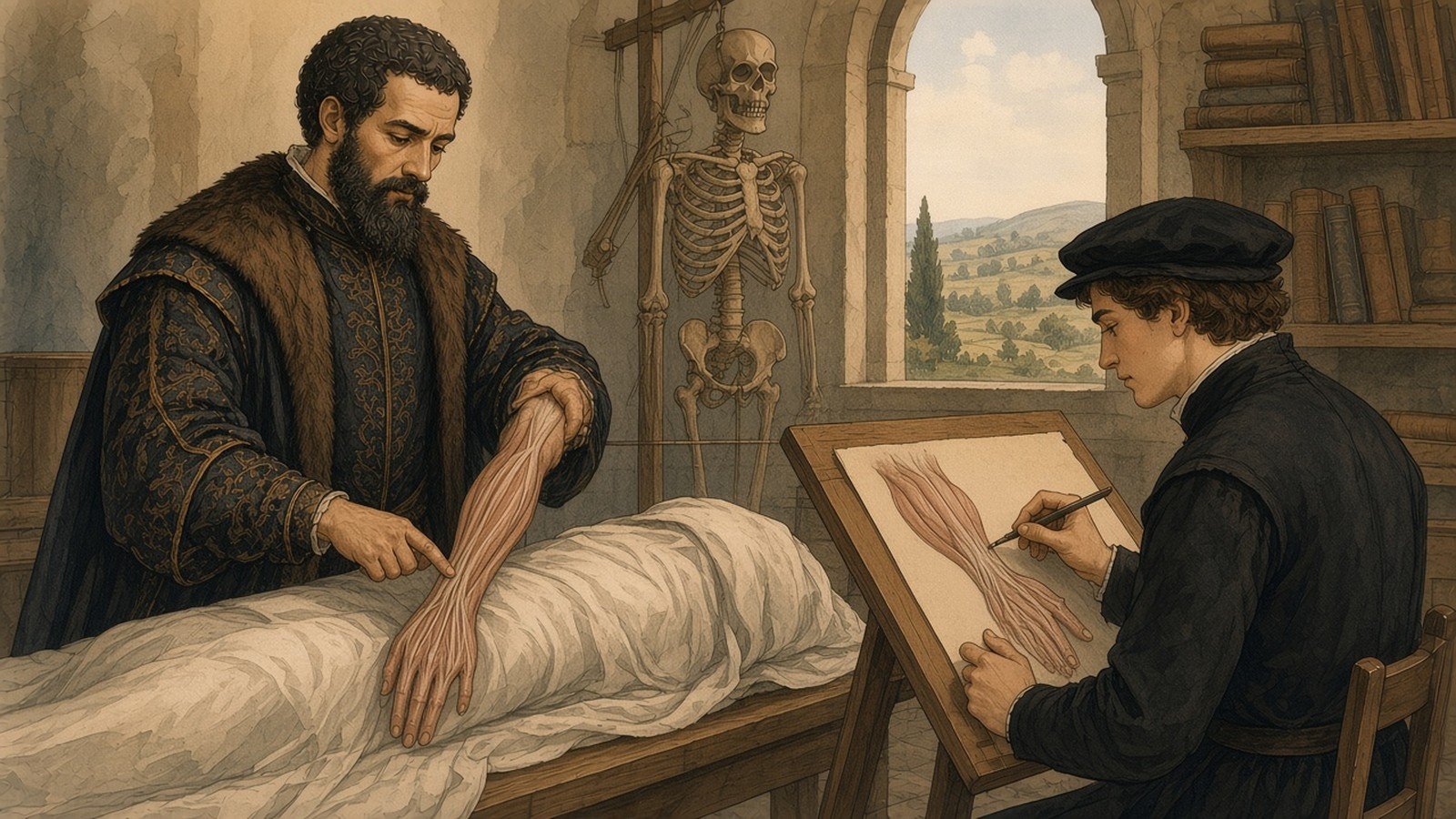
Inside the book is a portrait of Vesalius at the age of twenty-eight, the only likeness generally accepted as authentic. He stands beside a table, dressed in a fine gown, with a short beard and close-cropped hair, and holds up the dissected forearm and hand of a cadaver, displaying the tendons of the flexor muscles of the fingers. On the table lies a sheet of paper with a passage from the book on the muscles that move the fingers, and a pen and an inkpot. The choice of subject was deliberate. The hand was the instrument of the anatomist, and the dissection of the forearm was among the most difficult operations of the art. The portrait announced that the author had done the work with his own hands, and that the hand was itself an object of knowledge.
Seven Books
The seven books of the Fabrica follow an order of Vesalius’s own devising, which proceeds from the frame of the body inwards. The first book treats the bones and cartilages; the second, the ligaments and muscles; the third, the veins and arteries; the fourth, the nerves; the fifth, the organs of nutrition and generation in the abdomen; the sixth, the heart and the organs of the chest; and the seventh, the brain and the organs of sense. This arrangement differed both from the decay-driven order of Mondino and from the arrangement of Galen’s works, and it reflected Vesalius’s conviction that the bones were the foundation of the body, as walls and beams are the foundation of a house. At the end of the work is a chapter on the dissection of living animals, which described experiments on dogs and pigs to demonstrate the function of the nerves, the movement of the heart and lungs, and the action of the diaphragm.
The illustrations in the first two books are the most celebrated. The three full-page skeletons show the bones from different angles in poses that suggest life and thought rather than death: one leans on a spade, one rests its elbow on a tomb and gazes at a skull, and one raises its arms and seems to lament. The fourteen full-page “muscle men” show successive stages of a dissection in which the layers of muscle are progressively removed, from the surface down to the deepest layers, until in the last plates the figure, reduced nearly to the skeleton, hangs from a rope or leans against a wall. Each figure stands before a landscape of hills, villages and ruins. In the early twentieth century it was noticed that when the plates are laid side by side in a particular order the backgrounds join to form a continuous panorama, and later research identified the landscape with the Euganean Hills to the south-west of Padua, with the spa at Abano Terme recognisable among the buildings. The muscle men walk, gesture and turn as living bodies do, so that the reader sees how each muscle lies in the posture in which it acts.
The smaller woodcuts, set into the text, are no less important. They show individual bones from several sides, the instruments used in dissection, the stages of opening the skull and exposing the brain, and the branchings of the veins and nerves traced through the body. Each figure is lettered, and the letters are explained in a key beside the figure and referred to in the text, so that the reader could move between description and image with ease. The combination of exact text, keyed illustration and careful layout made the book a working tool as well as a monument, and it set a pattern for scientific illustration that persisted for centuries.
Galen Corrected
Vesalius did not set out to destroy Galen. He admired him, called him the prince of physicians, and followed him in a great deal, including in his physiology, the account of how the body worked. What he insisted on was that Galen’s anatomy should be judged by the body and not the body by Galen. In the preface to the Fabrica he wrote that Galen had been deceived by his apes, and in the body of the work he listed more than two hundred points on which Galen was wrong about human structure.
Many of these corrections concerned the bones. Galen, following the dog and the ape, had described the human lower jaw as made of two bones joined at the chin; Vesalius showed that in the adult human the mandible is a single bone. Galen described the sternum as having seven segments; Vesalius found three. He showed that the human breastbone, the hip bones and the sacrum differ from those of the ape, and he described the true number and form of the vertebrae. He showed that the human liver does not have five lobes, as in the dog, and that the human uterus is not divided into horns, as in many animals. He denied the existence in man of the rete mirabile, which had been a central element of Galenic physiology, the place where vital spirits were supposed to be refined into animal spirits on their way to the brain. Vesalius admitted that he himself had once taught its existence and had even shown it in his Tabulae, using the head of a sheep, and he wrote with some embarrassment of his former credulity.
On the heart, Vesalius was cautious in 1543. Galen had taught that blood passed from the right side of the heart to the left through invisible pores in the thick wall between the ventricles, the interventricular septum. Vesalius wrote that the pits on the surface of the septum did not, so far as he could see, pass through from one ventricle to the other, and that he was forced to marvel at the handiwork of the Creator who made blood pass through passages that escaped the sight. In the second edition of 1555 he went further, stating plainly that he could not see how even the smallest quantity of blood could pass through the substance of the septum. He did not, however, propose an alternative route, and he did not discover the circulation of the blood; that was left to his successors.
Not every correction was right, and Vesalius made errors of his own. He sometimes described animal structures as human when he lacked a suitable human specimen; one of his figures of the brain shows an ox’s structures, and his account of the female reproductive organs was influenced by Galen’s view that they were the male organs inverted. But the method he followed, and that he taught his readers to follow, was the means by which his own errors were later found and corrected. The Fabrica invited its readers to check every claim at the dissecting table, and many of them did.
Reception and Controversy
The book was admired at once for its beauty and attacked for its content. The most vehement attack came from Vesalius’s old teacher Sylvius in Paris, who in 1551 published a pamphlet entitled Vaesani cuiusdam calumniarum in Hippocratis Galenique rem anatomicam depulsio, “A refutation of the slanders of a certain madman against the anatomy of Hippocrates and Galen,” punning on Vesalius’s name. Sylvius argued that where the human body differed from Galen’s descriptions, the body had changed since antiquity; the human leg bones, for example, had become straight because modern men wore narrow trousers. Other critics were more measured but still loyal to Galen. Some colleagues at Padua resented the young professor’s confidence and the brilliance of his reputation.
Vesalius did not stay to fight these battles in the lecture theatre. Soon after the publication of the Fabrica he left Padua to become a physician to the household of Charles V, a post his family had held before him. The appointment was a great honour, but it effectively ended his career as a teacher and dissector. Before leaving he is said to have burned a large quantity of his notes and manuscripts, including a commentary on the Arabic physician Rhazes and annotated texts of Galen, in a fit of anger at the criticism of his work. He married Anne van Hamme in 1544 and followed the imperial court through Germany and the Low Countries, treating the emperor and his officers and gaining practical experience of surgery in war. In 1546 he published a letter on the medicinal properties of China root, a remedy then fashionable for the gout and syphilis, which included a long defence of his anatomical methods. In 1555 he brought out the second edition of the Fabrica, revised and corrected in many details and printed on heavier paper with a slightly larger type.
When Charles V abdicated in 1556, Vesalius was granted a pension and passed into the service of Philip II, whom he followed to Madrid in 1559. Spanish physicians were not welcoming, and he had little opportunity for anatomy, though he took part in the treatment of Philip’s son Don Carlos after a fall in 1562 that left the prince with a severe head injury. In 1561 the anatomist Gabriele Falloppio, who held Vesalius’s old chair at Padua, published his Observationes anatomicae, a respectful but critical commentary that corrected several of Vesalius’s statements from fresh dissections. Vesalius replied in 1564 with the Anatomicarum Gabrielis Falloppii observationum examen, conceding some points and defending others, and the exchange showed the new kind of anatomical argument that the Fabrica had made possible, in which professors disputed with one another about what they had seen in bodies rather than about what the ancients had written.
In 1564 Vesalius left Spain on a pilgrimage to Jerusalem. The reasons are obscure; later legends that he was condemned by the Inquisition for dissecting a living nobleman are unsupported by contemporary evidence. There are indications that he hoped to return to Padua, where Falloppio had died in 1562 and the chair was vacant. On the return voyage from the Holy Land his ship was caught in storms, and he fell ill. He was put ashore on the island of Zakynthos in the Ionian Sea, then a Venetian possession, and died there in October 1564, in his fiftieth year. The place of his grave is unknown.
The Paduan Succession
The most important consequence of the Fabrica was the school of anatomy that developed at Padua in the decades after Vesalius left. His immediate successor was Realdo Colombo, who had been his assistant and who, in his De re anatomica of 1559, described the passage of blood from the right ventricle of the heart through the lungs to the left side, the pulmonary transit, on the basis of vivisection; the Spanish theologian Michael Servetus had described the same route in a book of 1553 for which he was burned at Geneva, and the thirteenth-century Arabic physician Ibn al-Nafis had described it still earlier, though neither account was known to the anatomists of Padua. Falloppio, who followed Colombo, described the tubes leading from the ovaries to the uterus that bear his name, and much else besides. Falloppio’s pupil Girolamo Fabrici of Acquapendente held the chair for half a century, built the permanent anatomy theatre at Padua in 1594, which survives as the oldest of its kind, and described the valves in the veins in a treatise published in 1603.
Among Fabrici’s students at the end of the sixteenth century was a young Englishman, William Harvey, who took his medical degree at Padua in 1602. Harvey returned to London, and in 1628 he published Exercitatio anatomica de motu cordis et sanguinis in animalibus, in which he demonstrated, by a combination of dissection, vivisection, experiment and calculation, that the blood circulates continuously through the body, driven by the heart, out through the arteries and back through the veins. The valves that Fabrici had described were part of his evidence. Harvey’s argument rested on the habits of observation that Vesalius had taught, and in a real sense the line from the Fabrica to the circulation of the blood runs directly through the lecture theatre at Padua.
The influence of the Fabrica spread far beyond Padua. Its illustrations were copied, often without acknowledgement, by authors across Europe. Thomas Geminus in London published a plagiarised compendium in 1545 with copper engravings after the Vesalian woodcuts, the first anatomical copper plates in England. The Spanish anatomist Juan Valverde de Amusco published in 1556 a Spanish anatomy with plates closely based on those of Vesalius, which in its several editions and translations probably reached more readers than the Fabrica itself. Charles Estienne in Paris published his own illustrated anatomy, De dissectione partium corporis humani, in 1545, from blocks prepared earlier, and the comparison between the two books makes plain how far Vesalius had moved beyond his contemporaries. Through the seventeenth century the pattern of the illustrated anatomical atlas, exact, lettered and closely tied to the text, became the norm, and it survives in the anatomical atlases used by medical students today.
The Fate of the Woodblocks
The pearwood blocks from which the Fabrica and the Epitome were printed had a long and remarkable afterlife. After the second edition of 1555 they passed through the hands of several printers in Basel and Augsburg and were used for later editions and reprints. In the eighteenth century they were acquired by the Augsburg publishing house of Andreas Maschenbauer, and later they found their way to the university library at Ingolstadt, which moved to Landshut and then to Munich, where they were rediscovered in the 1890s. In 1934, in a project led by the New York Academy of Medicine and the University of Munich, the surviving blocks were used to print a magnificent folio, Andreae Vesalii Bruxellensis icones anatomicae, by the Bremer Presse in Munich, giving a new generation the chance to see impressions taken from the original woodcuts nearly four centuries after they were cut. The blocks were returned to the university library in Munich, and in July 1944 they were destroyed when Allied bombing set the library on fire. The 1934 printing thus became the last record of them.
The Fabrica itself has continued to be studied, translated and celebrated. A complete English translation by William Frank Richardson and John Burd Carman was published in five volumes between 1998 and 2009, and in 2014, for the five-hundredth anniversary of Vesalius’s birth, Daniel Garrison and Malcolm Hast published an annotated translation of both the 1543 and 1555 editions. The National Library of Medicine in Bethesda has made its copies and related works available in its online exhibition Historical Anatomies on the Web, which allows readers to page through the illustrations at high resolution.
Anatomy as a Science of Seeing
What made the Fabrica a turning point was not any single discovery. Many of its corrections of Galen were small matters of bones and lobes, and its physiology was still largely Galenic. Its importance lay in the method it embodied and the authority it claimed. Vesalius insisted that the anatomist must dissect with his own hands, that human anatomy must be learned from human bodies, that the evidence of the senses outranks the testimony of the ancients, and that knowledge of the body should be recorded in images exact enough to be checked by others. He combined in one volume the skills of the dissector, the scholar, the artist, the woodcutter and the printer, and he used the new technology of the press to make his observations reproducible across Europe. The book was published in the same year as Copernicus’s De revolutionibus orbium coelestium, and historians have often paired the two as the opening works of the scientific revolution. The pairing is apt in one respect: both books asked their readers to look again at things they thought they understood, and to trust what they could demonstrate over what they had been told.
The anatomy that Vesalius established became the foundation on which later medicine was built. Surgery depended on a precise knowledge of the course of vessels and nerves; the discovery of the circulation depended on an accurate knowledge of the heart and veins; pathology, as developed in the eighteenth and nineteenth centuries, depended on the comparison of diseased organs with a well-described normal anatomy. The surgeons who first operated under ether in the 1840s, described on this site in The Discovery of Anaesthesia, relied on an anatomy whose foundations were laid in the Fabrica, and when Wilhelm Röntgen’s X-rays first allowed physicians to see the bones of a living hand in 1895, the images they saw were read against the skeletons that Vesalius had drawn. The germ theory of disease, discussed in Pasteur, Koch and the Germ Theory of Disease, rested in turn on the careful comparison of healthy and diseased tissue that the anatomical tradition made possible.
The image of Vesalius that has come down to us is the one he chose for himself: a young man standing at a dissecting table, holding up the opened hand of a cadaver for the reader to see. In the history of anatomy that gesture marks the point at which the physician came down from the professor’s chair and began to look for himself, and at which the book became an instrument for showing what had been seen rather than a substitute for seeing it.