Antonie van Leeuwenhoek and the Discovery of Microbes
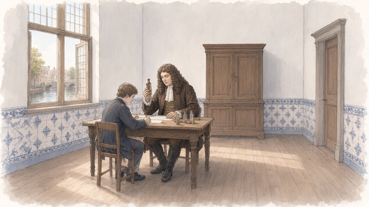
In the autumn of 1676 a draper and minor civic official of Delft in the Dutch Republic sent a long letter to the Royal Society in London describing what he had seen in drops of rainwater, well water, sea water and water in which peppercorns had been steeped. Using microscopes of his own making, each consisting of a single tiny lens mounted between two small metal plates, Antonie van Leeuwenhoek reported that the water was full of living creatures, which he called little animals, so small that many thousands of them could live in a single drop. Some of these were what are now called protists, single-celled organisms such as ciliates. Others, described in this and later letters, were bacteria. Leeuwenhoek was thus the first person known to have seen and described single-celled organisms, and in the course of half a century of observation he also gave early descriptions of spermatozoa, red blood cells, the flow of blood in capillaries and the structure of muscle fibres. This article describes Leeuwenhoek’s life and the world of Delft, the construction of his microscopes and why they performed so well, his principal discoveries and the way they were received in London, and the long interval before the organisms he described were connected with disease and with the chemistry of life.
A Draper of Delft
Antonie van Leeuwenhoek was born in Delft on 24 October 1632. His father, Philips Thoniszoon, was a basket maker, and his mother, Margaretha Jacobsdochter van den Berch, came from a family of brewers. The family name derived from the Lion’s Gate, the Leeuwenpoort, near which they lived. His father died when he was five, and his mother remarried. Leeuwenhoek was educated at a school at Warmond near Leiden and then lived for a time with an uncle at Benthuizen, who was an attorney and town clerk. At sixteen he was apprenticed to a linen draper in Amsterdam, where he worked as a bookkeeper and cashier.
In 1654 he returned to Delft, married Barbara de Mey, the daughter of a cloth merchant, and set up his own business as a draper, selling linen, ribbons and buttons. He bought a house on the Hippolytusbuurt, where he lived for the rest of his life. Of the five children of his first marriage, only one daughter, Maria, survived infancy. Barbara died in 1666, and in 1671 he married Cornelia Swalmius, the daughter of a clergyman, who died in 1694.
From 1660 Leeuwenhoek held a series of municipal offices that gave him a secure income independent of his trade. He was appointed chamberlain to the sheriffs of Delft, an office whose duties involved the upkeep of the room in the town hall in which the sheriffs met. In 1669 he qualified as a land surveyor, and in 1679 he was appointed wine gauger, responsible for checking the volume of wine imported into the city. These appointments, which he held for decades, gave him the leisure to pursue his researches.
Delft in the middle of the seventeenth century was a prosperous town of about twenty-five thousand people, known for its breweries, its pottery and its tapestry works. It was also the home of the painter Johannes Vermeer, who was born in the same month as Leeuwenhoek and who was baptised in the same year. When Vermeer died in 1675, Leeuwenhoek was appointed by the city as the trustee of his estate. Whether the two men knew each other well, and whether Leeuwenhoek was the model for Vermeer’s paintings of a geographer and an astronomer, have been suggested by some historians but cannot be established from the surviving evidence.
The Origins of Microscopy
Lenses for magnification had been used in Europe since the late thirteenth century, and simple magnifying glasses were common tools of the cloth trade, used by drapers to count the threads in a piece of fabric and judge its quality. Compound microscopes, combining two or more lenses in a tube, appeared in the Netherlands at about the same time as the telescope, in the first decades of the seventeenth century. Galileo, whose work on the telescope is described in the article on Galileo and the Telescope of 1609, adapted his instrument to observe small objects and described insects seen through it. In the 1620s and 1630s members of the Accademia dei Lincei in Rome published drawings of bees made with the aid of a microscope.
The work that brought microscopy to public attention was Micrographia, published in London in January 1665 by Robert Hooke, the curator of experiments of the Royal Society. Hooke used a compound microscope with an elaborate system of illumination and published large, detailed engravings of a flea, a louse, the eye of a fly, the point of a needle, the edge of a razor and many other objects. He described the small compartments in a thin slice of cork as cells, the first use of that word in biology. Micrographia was widely read, and it is likely that Leeuwenhoek knew of it, at least through its illustrations, since he is thought to have visited London around 1668. Some historians have suggested that Hooke’s description in the preface of how to make small spherical lenses from a thread of melted glass may have influenced Leeuwenhoek, though Leeuwenhoek’s own methods remained his secret.
The Single-Lens Microscope
Leeuwenhoek began making microscopes around 1670. His instruments were of a very different design from the compound microscopes of Hooke and other contemporaries. Each consisted of a single small lens, often less than a millimetre or two across and strongly curved, held in a hole between two thin rectangular plates of brass or silver about three to five centimetres long, riveted together. The specimen was mounted on the point of a pin in front of the lens. A system of screws allowed the pin to be moved up and down and towards or away from the lens, so that the specimen could be brought into position and focus. Liquid samples were placed in thin glass tubes or on small glass plates attached to the pin.
To use the instrument, the observer held it very close to the eye, with the plate almost touching the face, and looked through the lens towards a source of light, such as a window or a candle. The arrangement looks simple, almost crude, beside the elaborate tubes and stands of compound microscopes. It was nevertheless far superior in performance. Compound microscopes of the seventeenth century suffered badly from the aberrations of their lenses, which blurred and coloured the images, and the errors of each lens were multiplied by the next. A single lens, if ground or formed to a good shape and made small and strongly curved, could give a sharp image at high magnification.
Leeuwenhoek is known to have made more than five hundred lenses and several hundred microscopes, of which about ten survive with reasonable certainty of attribution. The best surviving instrument, in the collection of the University Museum in Utrecht, magnifies about 275 times and resolves details of about one micrometre, a thousandth of a millimetre. There is reason to think that Leeuwenhoek possessed instruments of greater power that he kept for his own use. He never revealed exactly how he made his best lenses. Some of the surviving lenses were ground and polished, and modern studies, including neutron tomography of surviving instruments carried out in 2018, have shown that at least some were ground rather than blown from glass. He also hinted in his letters at a method of observation that he would not disclose, which some historians have interpreted as a form of dark-field illumination, lighting the specimen from the side so that small objects appear bright against a dark background.
He made a separate microscope for many specimens, leaving the specimen permanently mounted, which explains in part the large number of instruments. At his death he left a cabinet of microscopes with specimens mounted, which was later sent to the Royal Society, as described below.
How a Single Lens Magnifies
The power of Leeuwenhoek’s instruments follows from simple optics. An object held close to the unaided eye cannot be seen clearly nearer than about twenty-five centimetres, the shortest distance at which a normal eye can focus. A converging lens placed immediately in front of the eye allows the object to be brought much closer while its image remains in focus, so that it fills a larger angle at the eye and appears larger. The magnification of such a simple microscope is roughly twenty-five centimetres divided by the focal length of the lens. A lens with a focal length of one millimetre therefore magnifies about two hundred and fifty times.
The focal length of a small glass sphere or strongly curved lens is proportional to its radius, so that the smaller the lens, the greater the magnification. The difficulty lies in making a lens only a millimetre or so across with surfaces accurate enough to form a sharp image, and in holding the specimen steady within a fraction of a millimetre of the correct distance. Leeuwenhoek’s screw adjustments solved the second problem, and his skill as a lens maker solved the first. The price of high magnification with a single lens is a very small field of view and a very short working distance, which made his microscopes awkward to use and demanded great patience in observation.
The compound microscope works differently. An objective lens forms an enlarged real image of the specimen inside the tube, and an eyepiece magnifies that image again. In principle this allows high magnification with lenses of moderate curvature, but in the seventeenth century each lens introduced spherical aberration, in which rays passing through different parts of a lens come to different foci, and chromatic aberration, in which light of different colours is focused at different points. In a compound instrument these errors were magnified by the eyepiece. Until achromatic objectives were developed in the nineteenth century, a well-made simple lens gave clearer images at high power than any compound microscope.
The Microscopes Today
The surviving instruments attributed to Leeuwenhoek are held in museums in the Netherlands, Belgium, Germany and the United States. Besides the Utrecht instrument, examples are held by the Rijksmuseum Boerhaave in Leiden, the Dutch national museum for the history of science and medicine. Several have been studied with modern methods. In 1981 the microbiologist Brian J. Ford examined specimens that Leeuwenhoek had sent to the Royal Society, which had been found among the Society’s papers, and photographed them through the Utrecht microscope, showing that it could resolve fine structures such as the cells of plant tissue and individual blood cells. His photographs demonstrated that Leeuwenhoek’s reports of what he could see were credible.
The neutron tomography study published in 2018 by Tiemen Cocquyt and colleagues of the Rijksmuseum Boerhaave and the Delft University of Technology examined the internal structure of several instruments without dismantling them. It found that the lenses examined differed in shape and quality, and that those studied had been made by grinding and polishing rather than by melting a bead of glass, as had sometimes been supposed. The study confirmed that Leeuwenhoek used conventional lens-making techniques with exceptional skill rather than any single secret process, though the methods of his very best lenses remain uncertain.
Contemporaries in Microscopy
Leeuwenhoek’s work belongs to a brief period in the second half of the seventeenth century in which a small number of observers transformed the study of living things with the microscope. Besides Hooke, the most important were Marcello Malpighi in Italy, Nehemiah Grew in England and Jan Swammerdam in the Netherlands.
Malpighi, a physician and professor at Bologna, described the capillaries in the lungs of a frog in 1661, studied the structure of the kidney, the spleen, the skin and the brain, and made detailed studies of the development of the chick embryo and of the anatomy of the silkworm. His studies of plant anatomy, published by the Royal Society in 1675 and 1679, appeared at the same time as those of Grew, a physician who published The Anatomy of Plants in 1682 and who first described the stomata of leaves and the structure of pollen grains in detail.
Jan Swammerdam of Amsterdam, a contemporary of Leeuwenhoek, studied the anatomy of insects with extraordinary care, dissecting them under the microscope with fine instruments of his own making. He showed that the metamorphosis of insects did not involve the sudden transformation of one creature into another but the gradual development of structures already present in the larva, and he gave one of the earliest descriptions of red blood cells, in the blood of a frog, in 1658. His great work on insects was published only in 1737 and 1738, long after his death in 1680, under the title Biblia Naturae, edited by Herman Boerhaave.
Unlike these men, who were trained physicians and anatomists, Leeuwenhoek did not dissect systematically or interpret his findings within a medical or anatomical framework. His strength lay in the power of his instruments and the range of his observations. After the deaths of Malpighi in 1694, Swammerdam in 1680 and Hooke in 1703, and with Leeuwenhoek’s refusal to train successors, microscopical research declined for much of the eighteenth century, a period some historians have described as a pause in the history of the microscope.
Leeuwenhoek’s own letters show that he did not regard himself as working in isolation. He read the Dutch translations of contemporary works when they were available, commented on the observations of others in his letters, and on several occasions corrected or disputed claims made by other microscopists, sometimes with considerable sharpness.
Correspondence with the Royal Society
Leeuwenhoek had little formal education. He knew no Latin, the language of learned communication, and could read and write only Dutch. He did not publish books in the usual sense. His discoveries became known through letters, about 190 of which he sent to the Royal Society in London over a period of fifty years, and others he sent to scholars and patrons in the Netherlands and elsewhere.
The connection was made by Regnier de Graaf, a physician of Delft known for his studies of the reproductive organs. In April 1673 De Graaf wrote to Henry Oldenburg, the secretary of the Royal Society, describing Leeuwenhoek’s microscopes as far better than any yet seen, and enclosed a letter from Leeuwenhoek describing observations of mould, the sting and mouthparts of a bee, and a louse. Oldenburg published an English translation in the Society’s journal, the Philosophical Transactions, and asked for more. From that time Leeuwenhoek wrote regularly. His letters were translated into English or Latin, often abridged, and published in the Philosophical Transactions, and they were later collected and published in Dutch and Latin editions in Leiden and Delft.
The letters were written in a conversational style, mixing careful description with accounts of his daily life, his methods of measurement and his reflections on what he saw. They were accompanied by drawings, which Leeuwenhoek did not make himself, since he said he could not draw well, but which were prepared by draughtsmen he employed and supervised.
The Little Animals
The most famous of Leeuwenhoek’s letters is dated 9 October 1676. In it he reported observations made since 1674 of water from a variety of sources. He had first noticed small creatures in the water of a lake, the Berkelse Mere near Delft, which became cloudy in summer. In rainwater collected in a new glazed pot and left to stand for some days, he found creatures of several kinds, some of them a thousand times smaller than the eye of a full-grown louse, moving through the water with wonderful agility. He described their shapes and movements, their manner of propulsion with small feet or threads, and their apparent numbers.
The most significant part of the letter concerned water in which he had soaked ground pepper, in an attempt to find out why pepper was pungent. In this infusion he found, among larger creatures, others so extraordinarily small that he estimated that more than a million of them would not equal in volume a grain of coarse sand. From the descriptions of their size and shape, historians and microbiologists have concluded that some of these were bacteria. Leeuwenhoek attempted to estimate their numbers by comparing their size with that of a grain of millet and calculating the number in a drop of water, and he reported that a single drop might contain more than two and a half million of them.
The claim was so extraordinary that the Royal Society did not at first accept it. When Hooke attempted in 1677 to reproduce the observations with his compound microscopes, he initially failed. Leeuwenhoek sent testimonials from eight reputable witnesses in Delft, among them a minister of the Reformed Church and a notary, who had looked through his microscopes and confirmed what he described. In November 1677 Hooke succeeded in seeing the small animals in pepper water with a single-lens microscope of his own, and demonstrated them to the Fellows of the Society, who signed a statement confirming that they had seen them. In 1680 Leeuwenhoek was elected a Fellow of the Royal Society. He never attended a meeting, but he remained deeply attached to the honour and continued to address his letters to the Society for the rest of his life.
Bacteria of the Mouth
Leeuwenhoek’s description of bacteria became clearer in a letter of 17 September 1683, in which he examined the white matter that collected between his teeth. Mixing it with clean rainwater and examining it under the microscope, he found many very small living animals, moving very prettily. He described several kinds, distinguished by their shapes and movements, and the drawings published with the letter show rods, spirals and spheres, corresponding to the principal shapes of bacteria now known as bacilli, spirilla and cocci. The figures in this letter are generally regarded as the first published illustrations of bacteria.
He extended the observations to the teeth of other people, among them two women, a child and an old man who had never cleaned his teeth in his life, and found differences in the kinds and numbers of organisms present. He observed that drinking very hot coffee killed the organisms on his front teeth but not those between his back teeth, and that vinegar killed them in a glass. These experiments, though informal, were among the first observations of the effect of heat and chemicals on microorganisms.
Spermatozoa and Generation
In 1677 Leeuwenhoek described spermatozoa, after a medical student at Leiden, Johan Ham, brought him a sample containing what Ham took to be animalcules. Leeuwenhoek examined human semen and that of many animals, including dogs, rabbits, fish, frogs and insects, and found such moving cells in all of them. He reported that they had a round body and a long tail, and that they were alive. In a letter to the Royal Society he took care to state that the human sample he examined had been obtained from conjugal intercourse and not by sinful means.
The discovery was taken up in a debate about generation, the question of how new organisms come into being. Leeuwenhoek argued that the spermatozoon was the essential agent of generation and that the egg, whose existence in mammals De Graaf had recently argued for, merely provided nourishment. This view, called animalculism, was opposed by ovism, which held that the embryo was contained in the egg. Some animalculists claimed to see a miniature human figure inside the head of the sperm, an image made famous by a drawing of 1694 by the Dutch microscopist Nicolaas Hartsoeker. Leeuwenhoek himself did not claim to have seen such a figure. The roles of sperm and egg were clarified only in the nineteenth century, when the fusion of the two cells in fertilisation was observed in sea urchins by Oscar Hertwig in 1876.
Leeuwenhoek opposed the doctrine of spontaneous generation, the belief that small animals such as insects, worms and the creatures in infusions arise from decaying matter. He studied the life cycles of fleas, weevils, ants, aphids and mussels and showed that they reproduced by eggs or by birth in the ordinary way. He found that aphids could reproduce without males, an early observation of parthenogenesis. The question of whether microorganisms could arise spontaneously remained open, however, and was settled only by the experiments of Louis Pasteur in the 1860s.
Blood, Muscle and Other Observations
The range of Leeuwenhoek’s observations was immense. In 1674 he gave one of the earliest descriptions of red blood cells, which he called globules, and measured their size by comparison with grains of sand; he observed that they were oval and nucleated in fish, frogs and birds, and round in mammals. In 1688 he observed the circulation of blood through the capillaries in the tail of a tadpole and in the fins of a small eel, confirming and extending the observation made by Marcello Malpighi in the lung of a frog in 1661. These observations completed the evidence for the circulation of the blood proposed by William Harvey in 1628, by showing how the blood passed from the arteries to the veins.
He described the striated structure of muscle fibres, the structure of the lens of the eye, the scales of the skin, the hairs of animals, the compound eyes of insects, the structure of wood and of ivory, the crystals in various salts, the structure of teeth and the spores of moulds. He described the single-celled alga Volvox and the green alga Spirogyra. In 1702 he described rotifers in the sediment of a gutter and observed that they could survive being dried out and revive when water was added, an early observation of the phenomenon now called anhydrobiosis. He observed protozoan parasites in his own stools, including organisms that have been identified as the intestinal parasite Giardia, during a bout of diarrhoea in 1681.
He also took a practical interest in measurement. Lacking any standard scale for small objects, he used comparisons with grains of sand, the diameter of a hair and the size of a red blood cell, and he devised methods of calculating volumes and numbers from these comparisons. His estimates of the sizes of the organisms he observed have been found to be generally consistent with modern measurements.
Visitors and Reputation
Leeuwenhoek’s discoveries made him a celebrity in his own lifetime. Many distinguished visitors came to his house in Delft to look through his microscopes, among them Queen Mary II of England, Gottfried Wilhelm Leibniz and, in 1698, Tsar Peter the Great of Russia, who was then travelling through the Netherlands and was shown the circulation of blood in the tail of an eel. Leeuwenhoek was reluctant to receive casual visitors, complaining that they took up his time, and he did not allow anyone to see his best instruments.
He refused to teach his methods to others or to take pupils, saying that most students wished only to make money or gain reputation and that few had the patience for such work. As a result, no one in his lifetime matched his skill, and for some decades after his death few observers could see what he had seen. The microscopes of the eighteenth century were mostly compound instruments used for the study of insects, plants and larger structures, and the systematic study of the smallest organisms advanced slowly.
Leeuwenhoek continued to observe and to write letters until the last weeks of his life. He died in Delft on 26 August 1723, at the age of ninety, and was buried in the Oude Kerk. His daughter Maria sent to the Royal Society, according to his wishes, a cabinet containing twenty-six of his microscopes, each with a specimen mounted. The cabinet was examined and described by Fellows of the Society but later disappeared, and its fate is unknown. The rest of his instruments were sold at auction in Delft in 1747.
From Animalcules to Microbiology
The connection between the organisms Leeuwenhoek described and the processes of disease, fermentation and decay was not established for more than a century and a half after his death. Throughout the eighteenth century the classification of microscopic organisms was confused. Carl Linnaeus, in his system of nature, grouped most of them in a single category, Chaos, among the worms, a treatment described in the article on Linnaeus and the System of Binomial Nomenclature. In 1786 the Danish naturalist Otto Friedrich Müller published the first systematic classification of microscopic organisms, including bacteria, and in 1838 Christian Gottfried Ehrenberg published a large work on infusoria.
The improvement of the compound microscope in the 1820s and 1830s, with achromatic objectives that corrected the colour fringes of earlier lenses, transformed the situation. Joseph Jackson Lister in London, the father of the surgeon Joseph Lister, did important work on the design of such objectives. With these instruments Matthias Schleiden and Theodor Schwann proposed in 1838 and 1839 that all plants and animals are composed of cells, and Rudolf Virchow argued in 1855 that every cell arises from a pre-existing cell.
In the 1850s and 1860s Louis Pasteur demonstrated that fermentation and putrefaction are caused by living microorganisms and that they do not arise spontaneously in sterile media protected from contamination. In the 1870s and 1880s Robert Koch identified the bacteria responsible for anthrax, tuberculosis and cholera and established methods for growing them in pure culture. The germ theory of disease that emerged from this work is described in the article on Pasteur, Koch and the Germ Theory of Disease. The bacteria that Leeuwenhoek had seen in pepper water and in the plaque of his teeth were now recognised as members of a vast kingdom of life, some of whose members caused disease, while most were harmless or essential to the processes of nature.
The study of the bacteria in the human mouth, which Leeuwenhoek had begun, was developed by Willoughby Dayton Miller in Berlin in the 1880s, who connected oral bacteria with tooth decay. The communities of bacteria living on and in the human body, now called the microbiome, have become a major field of research since the early twenty-first century, aided by methods of sequencing the genes of organisms that cannot be grown in the laboratory. The great majority of the species of bacteria and archaea in the world are now known chiefly through their genetic sequences rather than from cultures or microscopes.
Microbes and Medicine after Leeuwenhoek
The discoveries that followed from Leeuwenhoek’s observations made possible the control of infectious disease in the late nineteenth and twentieth centuries. Vaccination against smallpox had been introduced by Edward Jenner in 1796 without any knowledge of the causative agent, as described in the article on Edward Jenner and the Smallpox Vaccine, but the systematic development of vaccines against bacterial diseases followed from the work of Pasteur and Koch. Antiseptic surgery, introduced by Joseph Lister in the 1860s, rested on the recognition that infection of wounds was caused by microorganisms. The discovery of antibiotics, beginning with the observation of the effect of a mould on bacterial cultures by Alexander Fleming in 1928, told in the article on The Discovery of Penicillin by Alexander Fleming, provided the first general means of treating bacterial infections in the body.
Viruses, which are much smaller than bacteria and invisible in any light microscope, were recognised in the 1890s by Dmitri Ivanovsky and Martinus Beijerinck through their ability to pass through filters that retained bacteria, and they were first seen with the electron microscope in the 1930s. The resolving power of Leeuwenhoek’s best lenses, about one micrometre, was close to the limit set by the wavelength of visible light, which Ernst Abbe showed in 1873 to be about half the wavelength of the light used, or roughly two hundred nanometres. To see further required electrons instead of light.
Assessment
Leeuwenhoek’s achievement rested on a combination of technical skill and patient observation. He made instruments that, for their particular purpose, were better than any made by his learned contemporaries. He applied them over half a century to an extraordinary range of subjects, from the teeth of his neighbours to the eyes of insects, and he reported what he saw in detail, with careful estimates of size and number and with repeated observations under different conditions. He did not develop general theories, and some of his interpretations, notably his animalculist view of generation, were mistaken. But he opened a world of life that had been invisible, and his descriptions of single-celled organisms, of bacteria and of the fine structure of living tissues were the first steps towards microbiology, cell biology and histology. His position outside the universities and the learned societies, as a tradesman and municipal officer who wrote only in Dutch, makes his achievement the more notable, and his correspondence with the Royal Society shows how a scientific community could recognise and verify discoveries made by an observer with no formal standing.
Further Reading
The principal sources for this article include the Grokipedia entries linked in the text and the following: the digital edition of Leeuwenhoek’s letters, Alle de brieven van Antoni van Leeuwenhoek, published by the Digital Library for Dutch Literature; the collection of the University Museum Utrecht, which holds the most powerful surviving Leeuwenhoek microscope; and the account by Nick Lane of the 1676 letter, The unseen world, reflections on Leeuwenhoek (1677), in the Philosophical Transactions of the Royal Society B. The later history of microbiology is described in the article on Pasteur, Koch and the Germ Theory of Disease.