The Living Drug – CAR-T Cell Therapy
The Living Drug – CAR-T Cell Therapy
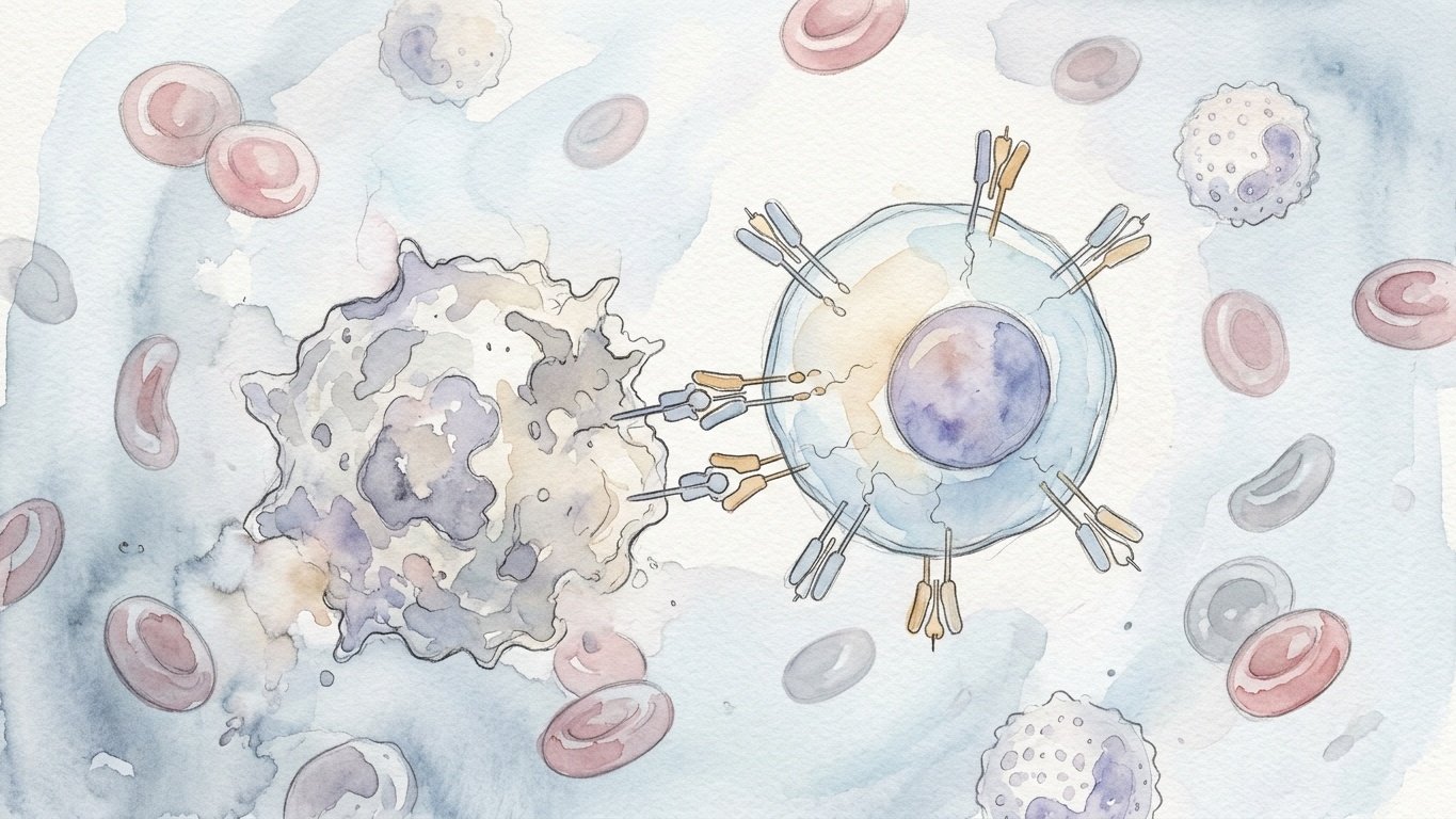
For more than a century, oncology was defined by three foundational pillars: the scalpel of the surgeon, the ionizing beam of the radiation oncologist, and the cytotoxic chemicals of systemic chemotherapy. Yet each of these modalities suffered from a tragic, intrinsic limitation: they were blunt instruments directed against biological processes that were profoundly dynamic, evolutionary, and evasive. Cancer cells are not foreign invaders with entirely alien biochemistry; they are corruptions of our own somatic self, mutating rapidly, downregulating surface markers, and erecting formidable biochemical microenvironments that disarm the host immune system. The dawn of chimeric antigen receptor (CAR) T-cell therapy represents the creation of a fundamentally new fourth pillar—the living drug. By extracting human cytotoxic T-lymphocytes from a patient’s bloodstream, genetically rewiring their intracellular signaling architecture with viral vectors, and infusing them back into the circulation as autonomous, self-replicating hunter-killer cells, bioengineers achieved what traditional pharmacology could never accomplish: a curative, intelligent therapeutic capable of clonal expansion, continuous target tracking, and decades-long immunological memory.
1. The Immunological Frontier and the Limits of Natural Surveillance
To understand why CAR-T cell therapy is regarded as one of the supreme achievements in biomedical engineering, one must first appreciate the staggering biological barriers that prevent the natural immune system from eradicating advanced malignancies. The human immune system possesses an extraordinarily sophisticated branch of adaptive immunity governed by cytotoxic CD8+ T-lymphocytes. In healthy physiology, these cellular sentinels circulate continuously through blood and lymphatics, scanning the surfaces of somatic cells for foreign peptides derived from intracellular pathogens or aberrant oncogenic proteins.
However, this surveillance mechanism is bound by an absolute molecular prerequisite known as Major Histocompatibility Complex (MHC) restriction, specifically human leukocyte antigen (HLA) Class I presentation. A conventional T-cell receptor (TCR) cannot directly recognize intact surface proteins. Instead, intracellular proteins must be degraded into short peptide fragments of eight to ten amino acids by the intracellular proteasome, transported into the endoplasmic reticulum by TAP transporters, loaded into the groove of an MHC Class I molecule, and exported to the plasma membrane. Only when a TCR engages both the foreign peptide and the self-MHC molecule with sufficient affinity—in concert with coreceptors and costimulatory signals—does the T-cell activate, polarize its cytoskeleton, and release lethal payloads of perforin and granzyme B into the immunological synapse, triggering apoptotic suicide in the target cell.
Malignant tumors exploit this strict dependency with devastating evolutionary efficiency. Under the selective pressure of immune surveillance—a phenomenon known as cancer immunoediting—tumor clones frequently lose expression of beta-2 microglobulin (B2M), mutate components of the antigen-processing machinery, or downregulate HLA Class I molecules altogether. By shedding their MHC presentation, cancer cells render themselves completely invisible to native T-cell receptors. Furthermore, malignant cells actively express inhibitory checkpoint ligands, such as Programmed Death-Ligand 1 (PD-L1), which bind to PD-1 receptors on infiltrating T-cells, transmitting biochemical signals that induce profound exhaustion, metabolic arrest, and apoptosis. Traditional monoclonal antibodies, while capable of binding intact surface antigens, act as passive pharmacological agents with fixed half-lives, lacking the autonomous ability to migrate into marrow sanctuaries, expand exponentially upon contact with target antigens, or persist as living immune memory. The biomedical challenge was therefore unprecedented: how could science endow cytotoxic T-cells with the antibody-like ability to recognize native, unmodified cell-surface antigens directly, entirely bypassing the requirement for MHC Class I presentation, while retaining the full proliferative, cytotoxic, and memory capabilities of a living lymphocyte?
2. The Architecture of the Chimeric Receptor – From Eshhar to Second-Generation Designs
The conceptual foundation of the chimeric antigen receptor was laid in the late 1980s by the Israeli immunologist Zelig Eshhar at the Weizmann Institute of Science in Rehovot. Working in collaboration with chemical immunologists, Eshhar asked whether the antigen-binding specificity of a monoclonal antibody could be genetically fused to the intracellular signaling domain of a T-cell receptor. The structural solution was elegant in its modularity, combining disparate functional domains from different branches of the immune system into a single synthetic fusion protein.
At the extracellular terminus, the receptor features a single-chain variable fragment (scFv). Constructed by taking the variable heavy (VH) and variable light (VL) chains of an antibody against a designated tumor-associated antigen and linking them via a flexible peptide linker (such as a pentapeptide repeat of Gly-Gly-Gly-Gly-Ser), the scFv preserves the exquisite target specificity and high nanomolar affinity of an antibody. This scFv domain is extended outward from the cell membrane by a flexible hinge or spacer domain, typically derived from immunoglobulin IgG subclasses (such as IgG1 or IgG4) or from CD8 alpha. The hinge provides structural flexibility and steric reach, allowing the scFv to bridge the spatial distance between the T-cell and the target epitope on the opposing plasma membrane.
Anchoring this extracellular construct is a hydrophobic transmembrane domain, frequently derived from CD28, CD8, or CD3 zeta, which traverses the phospholipid bilayer and connects the binding apparatus directly to the intracellular signaling machinery. In Eshhar’s original “first-generation” CAR design, the endodomain consisted exclusively of the intracellular signaling portion of the CD3 zeta chain, containing three Immunoreceptor Tyrosine-based Activation Motifs (ITAMs). When the scFv engaged its cognate antigen, receptor clustering triggered the phosphorylation of ITAM tyrosine residues by Src-family kinases such as Lck, creating docking sites for the tandem SH2 domains of ZAP-70, which in turn initiated the downstream calcium flux, MAPK cascade, and NFAT transcription factor translocation necessary for cytolytic degranulation.
Yet, when first-generation CAR-T cells were evaluated in early clinical trials against ovarian cancer, renal cell carcinoma, and lymphoma in the late 1990s and early 2000s, the clinical results were profoundly disappointing. The engineered cells demonstrated virtually no clinical efficacy. Within days of infusion, the circulating CAR-T cells disappeared from patients’ peripheral blood. In vitro assays revealed the fatal flaw: naive and memory T-cells require at least two distinct signals for sustained survival and clonal proliferation. Signal 1, delivered through the TCR/CD3 zeta complex, initiates activation; but in the absence of Signal 2—a costimulatory signal delivered through surface receptors such as CD28 or 4-1BB (CD137) upon interaction with ligands on mature antigen-presenting cells—Signal 1 drives the T-cell into an irreversible state of anergy or activation-induced cell death. First-generation CARs delivered an unmitigated Signal 1 without costimulation, causing the infused cells to exhaust and self-terminate upon their first encounters with tumor targets.
3. The Costimulatory Breakthrough – CD28 versus 4-1BB and Second-Generation Supremacy
The critical technological leap that transformed CAR-T from an academic curiosity into a life-saving therapy was the engineering of second-generation chimeric antigen receptors. In the late 1990s and early 2000s, independent research groups led by Michel Sadelain at Memorial Sloan Kettering Cancer Center in New York, Carl June at the University of Pennsylvania, and Malcolm Brenner at Baylor College of Medicine recognized that the intracellular domain must be built as a tandem signaling module. By cloning a costimulatory signaling domain in-series between the transmembrane region and the terminal CD3 zeta chain, bioengineers provided both Signal 1 and Signal 2 simultaneously upon a single engagement of the extracellular scFv.
Two primary costimulatory domains emerged at the forefront of clinical development, each conferring dramatically distinct biological, metabolic, and kinetic profiles upon the modified T-cells. Michel Sadelain championed the CD28 endodomain. Upon antigen binding, the CD28 domain recruits phosphatidylinositol 3-kinase (PI3K), triggering robust activation of the Akt and mTOR signaling pathways, which in turn stimulates rapid glycolytic metabolism, dramatic upregulation of anti-apoptotic proteins such as Bcl-xL, and copious secretion of interleukin-2 (IL-2). CD28-based CAR-T cells act like high-octane drag racers: they undergo explosive in vivo proliferation and execute ferocious, rapid cytolytic killing within 48 to 72 hours of infusion. However, this intense metabolic activation comes at the expense of longevity. CD28 CAR-T cells tend to differentiate rapidly into short-lived effector cells and suffer from earlier exhaustion, with persistence in patients typically measured in weeks or a few months.
Conversely, Carl June and his team at Penn, in collaboration with Dario Campana at St. Jude Children’s Research Hospital, focused on incorporating the intracellular signaling domain of 4-1BB (CD137), a member of the tumor necrosis factor receptor (TNFR) superfamily. Unlike CD28, 4-1BB signaling operates through the recruitment of TNFR-associated factors (TRAF1, TRAF2, and TRAF3), activating the non-canonical NF-kappa-B pathway and promoting mitochondrial biogenesis and oxidative phosphorylation rather than aerobic glycolysis. As a consequence, 4-1BB-costimulated CAR-T cells behave more like marathon runners: their initial cytolytic kinetics are slightly more gradual, but they exhibit profound resistance to exhaustion, maintain a central memory phenotype, and possess the remarkable capacity to persist in the human body for years—and in some patients, for more than a decade—acting as a permanent, living immunological shield against disease recurrence.
4. Choosing the Target – The Case of CD19 and B-Cell Malignancies
Even the most perfectly engineered chimeric receptor is useless without an ideal target antigen. The fundamental vulnerability of targeted cellular therapy is the risk of “on-target, off-tumor” toxicity: if the target antigen is expressed on vital healthy tissues—such as cardiac myocytes, hepatocytes, or pulmonary endothelium—the infused CAR-T cells will attack those organs with fatal consequences, as was tragically demonstrated in early trials targeting ERBB2 (HER2) with high-affinity receptors. The miraculous clinical success of the first commercial CAR-T therapies was made possible by the unique biology of a single cell-surface molecule: CD19.
CD19 is a 95-kilodalton transmembrane glycoprotein belonging to the immunoglobulin superfamily. It functions physiologically as a critical coreceptor on the surface of B-lymphocytes, complexing with CD21 (complement receptor 2), CD81, and CD225 to lower the threshold for B-cell receptor signaling and drive humoral immune responses. From an oncological perspective, CD19 possesses characteristics that approach perfection. It is expressed continuously throughout B-cell ontogeny, from the earliest committed pro-B cell stage through mature naive and memory B-cells, only being downregulated upon terminal differentiation into plasma cells. Crucially, CD19 is uniformly expressed on the vast majority of B-cell malignancies, including B-cell acute lymphoblastic leukemia (B-ALL), chronic lymphocytic leukemia (CLL), diffuse large B-cell lymphoma (DLBCL), and mantle cell lymphoma.
Equally vital is what CD19 does not express: it is entirely absent from pluripotent hematopoietic stem cells in the bone marrow, meaning that CAR-T ablation of CD19+ cells does not eradicate the patient’s underlying blood-forming capability. Furthermore, CD19 is absent from all non-hematopoietic tissues; it is not found in the brain, heart, lungs, kidneys, liver, or intestines. Targeting CD19 does indeed result in the complete eradication of healthy host B-cells—a collateral damage termed “B-cell aplasia.” However, humanity can survive indefinitely without mature B-lymphocytes, provided patients receive periodic prophylactic infusions of pooled intravenous immunoglobulin (IVIG) to supply protective antibodies against routine bacterial and viral infections. This biological window of dispensability made CD19 the proving ground upon which cellular immunotherapy would demonstrate its curative power.
5. The Trial of Courage – Carl June, Emily Whitehead, and the Battle Against Death
The turning point in the history of medicine arrived in the spring of 2012 at the Children’s Hospital of Philadelphia (CHOP). Six-year-old Emily Whitehead was dying of refractory pre-B acute lymphoblastic leukemia. Diagnosed at age five, she had undergone multiple rounds of aggressive cytotoxic chemotherapy, suffered devastating relapses, and had exhausted all conventional treatment protocols. Her bone marrow was packed with malignant leukemic blasts, and she had developed severe necrotic infections. Her physicians advised her parents to enter hospice care, estimating that she had mere days left to live.
In desperation, her parents enrolled her as the very first pediatric patient in a pioneering Phase I clinical trial run by oncologist Stephan Grupp at CHOP, utilizing an experimental 4-1BB/CD3-zeta anti-CD19 CAR-T construct developed across the street at the University of Pennsylvania by Carl June, David Porter, and Bruce Levine (designated CTL019). Emily’s autologous T-cells were harvested via leukapheresis, isolated, transduced with a replication-deficient lentiviral vector encoding the chimeric receptor, expanded over ten days in rocking bioreactor bags with anti-CD3/anti-CD28 magnetic Dynabeads, and infused into her veins over several days following conditioning chemotherapy with fludarabine and cyclophosphamide.
What followed was an unprecedented immunological storm that nearly cost Emily her life. Within days of the infusion, the engineered CAR-T cells encountered billions of CD19+ leukemic blasts in her bloodstream and bone marrow. The cells did not merely kill; they exploded in number, undergoing a thousand-fold clonal expansion inside her vascular system. As trillions of cytotoxic synapses formed and fired, the massive lysis of leukemic cells and the hyperactivation of host macrophages triggered a catastrophic systemic inflammatory syndrome. Emily developed raging fevers exceeding 105 degrees Fahrenheit, her blood pressure plummeted into profound refractory shock, her lungs filled with fluid requiring mechanical ventilation, and she entered multi-organ failure in the pediatric intensive care unit.
At that historic moment, clinical medicine had no protocol for this phenomenon, which is now universally known as Cytokine Release Syndrome (CRS). The clinical team faced a terrifying dilemma: if they administered high-dose corticosteroids to shut down the inflammatory cascade, they would likely kill the newly infused CAR-T cells and forfeit Emily’s only chance at surviving leukemia. Late one evening, Carl June examined emergency blood panels measuring twenty-four distinct cytokine levels in Emily’s serum. While many cytokines were elevated, one specific molecule stood out with astonishing prominence: Interleukin-6 (IL-6) was elevated to more than one thousand times above normal physiological levels.
In a flash of clinical brilliance, June remembered that his own daughter had recently been treated for juvenile idiopathic arthritis with a newly approved humanized monoclonal antibody named tocilizumab, which specifically blocks the IL-6 receptor (IL-6R). Tocilizumab had never been tested or administered for cytokine storm in an oncology patient. Grupp and the ICU team secured emergency off-label authorization and infused the drug directly into Emily’s intravenous line. The result was near-miraculous. Within hours, Emily’s vascular tone stabilized, her fevers broke, her arterial oxygenation improved, and within days she was successfully extubated. On Day 28 post-infusion, a repeat bone marrow biopsy revealed that her marrow was entirely free of leukemic blasts: she had achieved a complete, undetectable molecular remission. Today, Emily Whitehead remains cancer-free more than a decade later, serving as the global living symbol of the curative potential of cellular immunotherapy.
6. The Vector Engineering – Retroviruses, Lentiviruses, and Stable Integration
The manufacturing of a living cellular drug requires absolute precision in genetic delivery. Unlike small-molecule synthesis or recombinant protein production, where chemical reagents are reacted in stainless-steel vats, CAR-T manufacturing involves transforming a living, dying, metabolizing patient cell into a stable transgenic organism. Because T-lymphocytes undergo rapid cell division upon activation, transient gene delivery methods—such as electroporation of mRNA or non-integrating plasmid DNA—are insufficient for long-term therapy; the foreign genetic payload is diluted away with each round of mitosis, causing the cells to lose their chimeric receptors within days.
To achieve lifelong expression, bioengineers turned to retroviral and lentiviral vectors capable of permanently integrating the CAR transgene directly into the host T-cell’s chromosomal DNA. Early constructs utilized gamma-retroviruses derived from the Moloney murine leukemia virus (MoMLV). However, gamma-retroviruses suffer from two major biological drawbacks: they can only infect actively dividing cells (since they cannot traverse an intact nuclear envelope during interphase), and their integration pattern exhibits a dangerous bias toward transcription start sites and promoter regions of active genes, raising serious concerns regarding insertional mutagenesis and the potential activation of proto-oncogenes such as LMO2.
To overcome these hazards, modern clinical platforms—including the CTL019 platform that became Kymriah—adopted self-inactivating (SIN) lentiviral vectors derived from Human Immunodeficiency Virus type 1 (HIV-1). Through rigorous protein engineering, the pathogenic machinery of HIV is gutted. The viral genome is split across a four-plasmid packaging system: one plasmid encodes the Gag and Pol structural and enzymatic proteins; a second encodes the Rev regulatory protein required for nuclear export of unspliced viral RNA; a third encodes a heterologous envelope glycoprotein, most commonly the vesicular stomatitis virus G protein (VSV-G), which confers broad cellular tropism by binding to the universally expressed low-density lipoprotein (LDL) receptor; and the fourth transfer plasmid carries the chimeric antigen receptor expression cassette flanked by viral long terminal repeats (LTRs).
Crucially, the U3 region of the 3′ LTR is deleted in the transfer vector, creating a “self-inactivating” architecture. During reverse transcription inside the target T-cell, this deletion is copied to the 5′ LTR, entirely abolishing viral promoter and enhancer activity in the integrated provirus. Transcription of the CAR construct is instead driven exclusively by an internal, mammalian promoter—such as the human elongation factor-1 alpha (EF-1a) promoter or the cytomegalovirus (CMV) immediate-early promoter. Lentiviral vectors readily transduce both resting and activated T-cells by trafficking their pre-integration complexes through the nuclear pore complex via interactions with importin proteins, and their integration profile favors the introns of actively transcribed genes, vastly reducing the risk of leukemogenic proto-oncogene activation. The result is a safe, permanent genetic modification that is faithfully copied to every daughter cell as the T-cell clones divide.
7. The Manufacturing Crucible – Autologous Vein-to-Vein Logistics
The transition of CAR-T from an academic protocol to a standardized commercial pharmaceutical product required solving unprecedented logistical and bioengineering challenges. Conventional drugs are manufactured in centralized facilities, packaged into shelf-stable vials, and distributed through global wholesale networks to hospital pharmacies. Autologous CAR-T therapy, by contrast, is a bespoke “vein-to-vein” process where each batch of drug is manufactured exclusively for a single, designated human being.
The manufacturing cascade begins at the clinical treatment center with leukapheresis. The patient’s blood is routed through an apheresis centrifuge, which separates and collects the peripheral blood mononuclear cell (PBMC) fraction while returning red blood cells and granulocytes to the circulation. These raw PBMCs—which contain a chaotic mixture of CD4+ helper T-cells, CD8+ cytotoxic T-cells, B-cells, monocytes, natural killer cells, and often circulating malignant blasts—must be immediately stabilized, cryopreserved in dimethyl sulfoxide (DMSO), and shipped under strict liquid nitrogen vapor-phase dry vapor shippers (-150 degrees Celsius) to a specialized Good Manufacturing Practice (GMP) cellular processing facility.
Upon arrival at the central facility, the bag is thawed and subjected to rigorous cell selection. Monocytes are depleted, and T-lymphocytes are selected using magnetic bead sorting technology (such as anti-CD3/anti-CD28 magnetic microbeads). These beads do not merely isolate the cells; their surface-bound antibodies mimic physiological antigen presentation, delivering the simultaneous CD3 and CD28 signals required to jolt resting T-cells out of G0 arrest and drive them into rapid cell division. Once the cells are actively cycling, they are transduced with the clinical-grade lentiviral or retroviral vector at a defined multiplicity of infection (MOI).
The transduced cells are then transferred into automated, closed-system bioreactors—such as the Miltenyi CliniMACS Prodigy or rocking-motion wave bioreactor bags. Over seven to fourteen days, the cells are cultivated in serum-free medium fortified with recombinant human cytokines, predominantly Interleukin-7 (IL-7) and Interleukin-15 (IL-15), which favor the expansion of early-lineage stem cell memory (Tscm) and central memory (Tcm) subsets over terminal effector phenotypes. When the cell harvest reaches hundreds of millions or billions of viable CAR-expressing cells, the magnetic activation beads are magnetically stripped away, the cells are washed, concentrated, formulated into infusion bags with cryoprotectant, and subjected to exhaustive quality control testing. Every single patient lot must pass strict release criteria: sterility tests for bacterial, fungal, and mycoplasma contamination; endotoxin levels; lentivirus replication-competent lentivirus (RCL) assays; cell viability (>80%); and vector copy number (VCN) quantification via droplet digital PCR to ensure that average proviral insertions remain safely below five copies per genome to prevent insertional oncogenesis. Only after clearing this gauntlet is the personalized cellular product frozen, shipped back across continents to the patient’s bedside, thawed in a water bath, and infused over thirty minutes.
8. Clinical Pathophysiology – Cytokine Release Syndrome and ICANS
As CAR-T clinical trials expanded globally from leukemia to aggressive B-cell lymphomas, the medical community confronted the dark side of living cellular therapeutics: acute, life-threatening toxicities directly driven by immunological success. The two primary toxicities—Cytokine Release Syndrome (CRS) and Immune Effector Cell-Associated Neurotoxicity Syndrome (ICANS)—represent distinct pathophysiological entities that require rigorous clinical grading and algorithmic intervention.
Cytokine Release Syndrome is an acute systemic inflammatory response initiated by the massive activation and proliferation of CAR-T cells upon binding their target antigens. When millions of CAR-T cells simultaneously engage tumor cells, they release copious quantities of interferon-gamma (IFN-g) and granulocyte-macrophage colony-stimulating factor (GM-CSF). These effector cytokines do not act alone; they serve as a powerful siren call that recruits and hyperactivates bystander myeloid cells, particularly host monocytes and macrophages. These activated macrophages, in turn, become the primary cytokine factories, unleashing massive systemic waves of Interleukin-6 (IL-6), Interleukin-1 (IL-1), tumor necrosis factor-alpha (TNF-a), and inducible nitric oxide synthase (iNOS).
The resulting hypercytokinemia leads to widespread endothelial cell activation, capillary leak syndrome, and profound systemic vasodilation. Clinically, CRS manifests along a spectrum: Grade 1 presents with isolated high fevers; Grade 2 involves moderate hypotension responsive to fluids and mild hypoxia requiring low-flow nasal cannula oxygen; Grade 3 escalates to severe hypotension requiring high-dose vasopressors (such as norepinephrine) and high-flow oxygen; and Grade 4 represents life-threatening shock, acute respiratory distress syndrome (ARDS) requiring mechanical ventilation, and multi-organ failure. The management of CRS was revolutionized by the standardization of the anti-IL-6 receptor antagonist tocilizumab, often combined with the IL-1 receptor antagonist anakinra and systemic corticosteroids (dexamethasone or methylprednisolone) for refractory cases, transforming what was once a 30% mortality event into a manageable clinical complication.
Immune Effector Cell-Associated Neurotoxicity Syndrome (ICANS), by contrast, represents a profound central nervous system disturbance that typically manifests several days after the peak of CRS. Patients experience expressive aphasia, word-finding difficulty, tremors, lethargy, global confusion, and in severe cases, seizures, motor weakness, cerebral edema, and coma. The underlying mechanism does not appear to involve direct infiltration of CAR-T cells attacking brain parenchyma, but rather severe endothelial breakdown at the blood-brain barrier. High systemic levels of inflammatory cytokines, particularly IL-6, IL-1, and angiopoietin-2, disrupt endothelial tight junctions in cerebral microvessels, allowing systemic cytokines, myeloid cells, and protein-rich fluid to pour into the cerebrospinal fluid and brain extracellular space. Interestingly, tocilizumab is generally ineffective against ICANS—and can occasionally worsen it—because the antibody does not cross the blood-brain barrier, while blocking peripheral IL-6 receptors can transiently increase circulating levels of free IL-6 in the serum, which then crosses the compromised barrier into the brain. Consequently, high-dose corticosteroids, which readily penetrate the central nervous system, and IL-1 antagonists remain the gold standard for halting neurotoxic progression.
9. Regulatory Milestones and the Commercial Landscape
The dramatic clinical remissions observed in clinical trials—where patients with terminal, chemo-refractory leukemia and diffuse large B-cell lymphoma who had 6-month survival expectations of less than 10% were suddenly achieving complete remission rates of 50% to 80%—forced a revolution within regulatory agencies. In August 2017, the United States Food and Drug Administration (FDA) made medical history by granting landmark approval to tisagenlecleucel (marketed by Novartis as Kymriah) for the treatment of pediatric and young adult patients with relapsed or refractory B-cell acute lymphoblastic leukemia. It was the first gene therapy and the first cellular therapy ever approved in the Western hemisphere.
Just two months later, in October 2017, the FDA approved axicabtagene ciloleucel (marketed by Kite Pharma/Gilead as Yescarta), a CD28-costimulated CAR-T therapy for adult patients with relapsed or refractory diffuse large B-cell lymphoma (DLBCL) following two or more lines of systemic therapy. These historic approvals were rapidly followed by a wave of second-generation CAR-T authorizations that expanded the therapeutic beachhead across hematological oncology:
- Brexucabtagene autoleucel (Tecartus): Approved in 2020 for relapsed or refractory mantle cell lymphoma (MCL) and adult B-ALL, utilizing a manufacturing process that removes circulating CD19+ leukemic blasts during processing to prevent premature CAR-T cell activation and exhaustion.
- Lisocabtagene maraleucel (Breyanzi): Approved in 2021 for large B-cell lymphoma, featuring a precisely defined 1:1 ratio of CD4+ helper and CD8+ cytotoxic CAR-T cells to reduce pharmacokinetic variability and decrease the incidence of severe CRS and neurotoxicity.
- Idecabtagene vicleucel (Abecma) and Ciltacabtagene autoleucel (Carvykti): Approved in 2021 and 2022, marking the historic expansion of CAR-T beyond CD19 into B-cell maturation antigen (BCMA) for the treatment of relapsed and refractory multiple myeloma. Carvykti, utilizing two distinct heavy-chain-only variable domains targeting two discrete epitopes on BCMA, achieved unprecedented overall response rates exceeding 97% in heavily pretreated myeloma patients.
These clinical triumphs fundamentally altered the oncology standard of care, moving CAR-T from third-line salvage therapy into second-line treatment, directly competing with and outperforming autologous stem cell transplantation in major randomized Phase III clinical trials such as ZUMA-7 and TRANSFORM.
10. The Mechanisms of Tumor Resistance and Antigen Escape
Despite the unprecedented response rates of CAR-T therapy, oncology remains locked in an evolutionary arms race with the malignant genome. Long-term follow-up studies reveal that approximately 40% to 60% of patients who achieve initial complete remission eventually relapse. Understanding the precise molecular mechanisms of resistance has become the foremost priority of contemporary immunotherapy research.
Resistance mechanisms divide into two major categories: antigen-positive relapse and antigen-negative escape. In antigen-positive relapse, the tumor cells continue to express robust levels of the target antigen (such as CD19), but the infused CAR-T cells fail to persist or undergo premature exhaustion. Single-cell RNA sequencing of exhausted CAR-T cells reveals the persistent upregulation of multiple inhibitory receptors, including PD-1, TIM-3, LAG-3, and TIGIT, accompanied by a profound loss of proliferative capacity and cytotoxic cytokine secretion. Epigenetic profiling has shown that exhausted T-cells undergo stable chromatin remodeling, with de novo methylation and silencing of critical memory and stem-like transcription factors such as TCF-1 (encoded by TCF7), locking the cells into an irreversible state of dysfunction.
Antigen-negative relapse, by contrast, represents a masterpiece of Darwinian selection under immunological pressure. Confronted with a living drug that systematically destroys any cell bearing CD19, tumor populations emerge that have completely lost surface antigen expression. In B-ALL, researchers discovered that malignant cells evade anti-CD19 CARs through diverse genetic and post-transcriptional stratagems:
First, tumors frequently acquire frameshift mutations or deletions in the CD19 gene on chromosome 16, resulting in nonsense-mediated mRNA decay or truncated proteins that fail to localize to the plasma membrane. Second, alternative splicing plays a insidious role: tumors frequently upregulate a splice variant that skips exon 2 of CD19 (CD19-Delta-exon-2), which encodes the exact extracellular epitope recognized by the FMC63 antibody clone used in Kymriah and Yescarta; the truncated CD19 protein continues to signal internally, sustaining leukemic survival, while completely evading CAR detection. Third, in rare and devastating cases, leukemic cells undergo lineage switching: under the selective pressure of CD19 CAR-T, B-ALL clones undergo phenotypic transdifferentiation into acute myeloid leukemia (AML), shedding all B-lymphoid markers including CD19, CD20, and CD22, and adopting a myeloid morphology and expression profile (CD33, CD123) that leaves the engineered T-cells helpless.
11. Overcoming the Solid Tumor Fortress – The Microenvironmental Siege
While CAR-T cell therapy has revolutionized hematological malignancies, its translation to solid tumors—which account for more than 90% of all cancer deaths, including carcinomas of the lung, breast, prostate, colon, and pancreas—has proven to be an extraordinarily difficult biological challenge. The reasons for this disparity lie in the physical and immunological architecture of solid neoplasms.
The first obstacle is trafficking and infiltration. In hematological cancers, CAR-T cells and target malignant cells mingle freely in the liquid environments of peripheral blood, spleen, and bone marrow. In solid tumors, the engineered cells must extravasate from tumor microvessels, migrate across dense extracellular stroma composed of crosslinked collagen and hyaluronic acid, and penetrate deep into hypovascular, necrotic tumor cores. Many solid tumors express mismatched chemokines or downregulate adhesion molecules (such as ICAM-1 and VCAM-1), effectively barricading their tissue borders against lymphocyte entry.
The second obstacle is antigen heterogeneity. Unlike B-cell leukemias, where CD19 is clonal and expressed across 100% of cells, solid tumors exhibit staggering intratumoral genetic and phenotypic heterogeneity. Antigens such as EGFRvIII in glioblastoma, mesothelin in pancreatic adenocarcinoma, or HER2 in breast cancer are frequently expressed on only a subpopulation of tumor cells. CAR-T cells targeting a single antigen may eradicate 70% of the tumor mass, only for the antigen-negative clones to rapidly outgrow and kill the host.
The third and most formidable obstacle is the immunosuppressive tumor microenvironment (TME). Solid tumors are surrounded by a hostile metabolic and immunological fortress. The metabolic milieu is characterized by extreme hypoxia, severe acidosis (low pH due to lactic acid accumulation from the Warburg effect), nutrient starvation (depletion of glucose, glutamine, and essential amino acids like tryptophan by indoleamine 2,3-dioxygenase, IDO), and high concentrations of immunosuppressive adenosine. Immunologically, the tumor recruits regulatory T-cells (Tregs), myeloid-derived suppressor cells (MDSCs), and tumor-associated M2 macrophages, which secrete paralyzing concentrations of transforming growth factor-beta (TGF-b) and Interleukin-10 (IL-10). When a standard second-generation CAR-T cell enters this toxic environment, its mitochondrial machinery fails, its cytotoxic machinery is extinguished, and it rapidly enters terminal exhaustion.
12. Fourth-Generation TRUCKs, Logic Gates, and the Synthetic Biology Revolution
To breach the solid tumor fortress, synthetic immunologists are designing fourth- and fifth-generation CAR architectures, transforming T-cells from simple targeted killers into autonomous microscopic pharmaceutical factories and computational biological devices.
One major innovation is the development of “TRUCKs” (T-cells Redirected for Universal Cytokine-mediated Killing), also designated fourth-generation CARs. These cells are engineered with an inducible transgenic expression cassette under the control of an NFAT-responsive promoter. When the CAR binds its target antigen, the resulting calcium-NFAT signaling cascade not only triggers cytolysis, but also induces the localized secretion of potent pro-inflammatory cytokines, such as Interleukin-12 (IL-12), Interleukin-18 (IL-18), or Interleukin-7/IL-15 fusions. By depositing IL-12 directly into the tumor bed, the engineered T-cell remodels the local stroma, repolarizes suppressive M2 macrophages into tumor-killing M1 phenotypes, recruits host natural killer (NK) cells, and eliminates surrounding antigen-negative tumor variants through bystander killing without causing systemic cytokine toxicity.
Simultaneously, synthetic biology has introduced computational logic gates to solve the challenge of tumor specificity and avoid lethal on-target off-tumor toxicities. Using synthetic Notch (synNotch) receptors developed by Wendell Lim at the University of California, San Francisco, bioengineers can construct “AND” gates: a T-cell is engineered with a synNotch receptor that recognizes Antigen A; upon engagement, synNotch cleaves an intracellular transcription factor that translocates to the nucleus to drive the de novo expression of a CAR targeting Antigen B. Thus, the T-cell only activates and destroys a target cell if both Antigen A and Antigen B are present simultaneously on the same tissue, sparing healthy organs that express only one of the antigens. Conversely, “NOT” gates utilize inhibitory chimeric antigen receptors (iCARs) equipped with intracellular signaling domains from PD-1 or CTLA-4. If the T-cell encounters a target antigen on healthy tissue that also expresses a designated normal tissue marker, the iCAR delivers an overriding negative signal that aborts activation, protecting normal cells while permitting the destruction of tumor cells that have lost the normal marker.
13. Allogeneic “Off-the-Shelf” CAR-T – CRISPR and Universal Donor Engineering
While autologous CAR-T therapy has saved thousands of lives, its widespread global adoption is constrained by immense economic, logistical, and biological hurdles. The autologous manufacturing process takes two to four weeks, costs between $375,000 and475,000 per dose for the cellular product alone, and occasionally fails due to poor T-cell quality in heavily pretreated, lymphopenic patients. Patients with hyper-aggressive leukemias or rapidly progressing lymphomas frequently deteriorate and die before their personalized cells can be manufactured and released. The holy grail of cellular oncology is therefore the creation of allogeneic, “off-the-shelf” CAR-T therapies: pre-manufactured batches of universal donor T-cells derived from healthy volunteers, cryopreserved in thousands of identical doses, and immediately available for infusion upon clinical demand.
However, infusing allogeneic T-cells from a healthy donor into an HLA-mismatched patient triggers two devastating immunological catastrophes: Graft-versus-Host Disease (GvHD) and rapid host rejection. GvHD occurs when the donor T-cells’ endogenous alpha-beta T-cell receptors recognize the recipient’s normal tissues as foreign, launching an attack on the skin, liver, and gastrointestinal tract. Rejection occurs when the host’s surviving immune system recognizes the foreign donor cells’ HLA molecules and destroys them before they can eradicate the tumor.
To overcome these barriers, scientists are deploying advanced multiplex gene editing using CRISPR-Cas9, transcription activator-like effector nucleases (TALENs), and base editing. To eliminate GvHD, researchers genetically knock out the TRAC gene (T-cell receptor alpha constant region), permanently deleting the native alpha-beta TCR from the cell surface. To prevent host rejection, the beta-2 microglobulin (B2M) gene is knocked out, eliminating HLA Class I surface expression, often accompanied by the targeted insertion or overexpression of non-classical HLA molecules (such as HLA-E or CD47) to prevent recipient Natural Killer (NK) cells from killing the “missing-self” donor cells. Pioneered in clinical trials by Cellectis, Allogene Therapeutics, and academic centers worldwide, universal allogeneic CAR-T cells have already demonstrated proof-of-concept remissions, heralding an era where curative cellular therapies will be mass-produced with consistent pharmaceutical quality and distributed globally at a fraction of current costs.
14. Beyond Oncology – Autoimmunity, Fibrosis, and Senescence
The triumph of CAR-T cell engineering in cancer has illuminated an even broader biological horizon: the application of living therapeutics to non-malignant human diseases. Because chimeric receptors can be designed to target virtually any cell-surface protein, the platform is rapidly expanding into rheumatology, cardiology, and regenerative medicine.
The most spectacular breakthrough has occurred in autoimmune diseases, led by Georg Schett and his team at the University of Erlangen-Nuremberg in Germany. In systemic lupus erythematosus (SLE), idiopathic inflammatory myositis, and systemic sclerosis, autoantibodies produced by autoreactive B-cells drive catastrophic multi-organ inflammation and tissue destruction. In landmark clinical studies published in 2022 and 2024, patients with severe, life-threatening refractory lupus were treated with autologous CD19-directed CAR-T cells. The engineered cells swept through the patients’ blood, lymph nodes, and tissues, completely obliterating the pathogenic B-cell compartment. Following B-cell aplasia, the patients achieved complete drug-free remissions, autoantibodies disappeared entirely, and proteinuria resolved. Crucially, months later, naive B-cells re-emerged from the bone marrow hematopoietic stem cells without autoreactivity: the living drug had successfully executed an immunological “hard reboot,” resetting the immune system to a healthy, tolerant baseline.
In cardiology, researchers at the University of Pennsylvania led by Jonathan Epstein have engineered CAR-T cells targeting Fibroblast Activation Protein (FAP). In murine models of hypertensive heart disease and cardiac hypertrophy, activated cardiac fibroblasts secrete excessive extracellular matrix, causing myocardial fibrosis and heart failure. Infusion of anti-FAP CAR-T cells selectively ablated the activated fibroblasts, dramatically reducing cardiac fibrosis and restoring systolic and diastolic ventricular function. In 2022, the same group demonstrated that transient CAR-T cells could be generated in vivo inside the patient’s body by injecting lipid nanoparticles (LNPs) carrying modified mRNA encoding an anti-FAP CAR targeted to T-cells via surface-conjugated anti-CD5 antibodies, eliminating the entire ex vivo manufacturing infrastructure.
Finally, in the biology of aging, cellular senescence has emerged as a central driver of chronic inflammation and tissue degeneration. Senescent cells enter permanent cell cycle arrest, resist apoptosis, and secrete a toxic cocktail of pro-inflammatory factors known as the senescence-associated secretory phenotype (SASP). By designing CAR-T cells targeting urokinase-type plasminogen activator receptor (uPAR)—a surface marker specifically upregulated on senescent cells—researchers at Memorial Sloan Kettering demonstrated that senolytic CAR-T cells could selectively eliminate senescent cells in aging mice, reversing hepatic fibrosis, improving exercise capacity, and extending healthy lifespan. The living drug has evolved into an engine of biological renewal.
15. The Living Drug and the Future of Medicine
The journey of CAR-T cell therapy—from Zelig Eshhar’s initial recombinant experiments in Rehovot, to the pioneering protein engineering of Michel Sadelain and Malcolm Brenner, to the dramatic clinical breakthrough of Carl June and Emily Whitehead at Penn, to the modern frontiers of multiplex CRISPR editing and in vivo LNP programming—represents one of the supreme intellectual triumphs in the history of science. It marks the definitive transition of medicine from the paradigm of passive chemistry to that of active, synthetic biology.
For millennia, human therapeutics consisted of small molecules and static proteins: compounds that obeyed classical pharmacokinetics, circulating through plasma, binding passively to receptors, degrading over hours, and possessing zero awareness of their biological environment. CAR-T cell therapy replaced that paradigm with an intelligent, autonomous entity. A single dose of a few million cells contains within its genetic code the capacity to seek out microscopic disease across an entire human anatomy, expand a thousand-fold into a personalized army in response to antigen contact, orchestrate complex cellular dialogues with surrounding immune cells, dismantle pathological tissues, and establish permanent sentinels that persist for decades to guard against recurrence.
As we stand in the mid-2020s, the challenges that remain—penetrating the immunosuppressive armor of solid tumors, manufacturing universal allogeneic cells at commercial scale, and expanding into autoimmunity and cardiovascular disease—are being dismantled by the convergence of synthetic biology, computational immunology, and genome engineering. CAR-T cell therapy has proven that the human immune system, when re-architected by scientific intellect, is the most lethal, precise, and curative weapon ever deployed against human disease. The era of the living drug has arrived, and it has forever altered the destiny of human health.