Mind and Silicon – Intracortical BCIs
Mind and Silicon – Intracortical BCIs
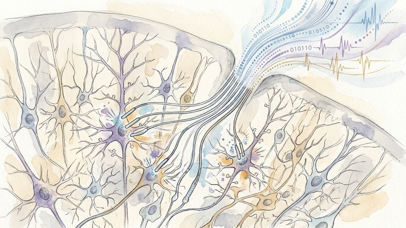
Inside the human cranium lies the most complex physical structure in the known universe: eighty-six billion neurons interconnected by more than one hundred trillion synaptic junctions, computing thoughts, memories, emotions, and motor intentions through rapid millivolt electrochemical action potentials. For centuries, this internal symphony of consciousness was fundamentally locked behind the biological barrier of the skull, accessible only through the physical execution of peripheral nerves and skeletal muscles. When trauma, stroke, or neurodegenerative diseases—such as Amyotrophic Lateral Sclerosis (ALS), cervical spinal cord injury, or brainstem stroke—sever these motor pathways, the conscious mind remains intact, trapped in a paralyzed body in the terrifying state known as ‘locked-in syndrome.’ The development of high-density intracortical Brain-Computer Interfaces (BCIs) represents the ultimate convergence of neurophysiology, microelectronics, materials science, and machine learning: the direct physical coupling of biological neural tissue with digital silicon. By transitioning from rigid, 96-channel silicon needles to thousands of micron-thin flexible polymer electrode threads inserted by robotic microsurgery, and decoding real-time neural population vectors with recurrent neural networks and transformer models, bioengineers have shattered the skull’s isolation. Today, locked-in patients are controlling digital interfaces, commanding high-degree-of-freedom robotic limbs, and communicating at natural conversational speeds purely through the electrical firing of their cortical thoughts.
1. The Electrical Mind – From Galvani to the Hodgkin-Huxley Landscape
The scientific realization that the mind operates via electrical signals began in the late eighteenth century with Luigi Galvani’s experiments in Bologna, demonstrating that bioelectric currents animate muscular contraction. In 1875, Richard Caton recorded electrical fluctuations from the cerebral cortex of rabbits and monkeys, paving the way for Hans Berger’s landmark 1924 invention of the electroencephalogram (EEG) in Jena, Germany, capturing the synchronized rhythmic oscillations of human brain waves through the intact scalp.
The definitive biophysical description of neural signaling was formulated in 1952 by Alan Hodgkin and Andrew Huxley at Cambridge University, earning them the 1963 Nobel Prize in Physiology or Medicine. Working on the giant axon of the Atlantic squid (Loligo pealeii), Hodgkin and Huxley established that a neural action potential is a transient, self-propagating electrical wave generated by the voltage-dependent opening and closing of microscopic ion channels across the neuronal plasma membrane.
In the resting state, ATP-driven sodium-potassium pumps (Na⁺/K⁺ ATPase) maintain an asymmetric distribution of ions, establishing a negative resting membrane potential of approximately -70 millivolts inside the neuron. When synaptic inputs depolarize the membrane to its threshold (approximately -55 mV), voltage-gated sodium channels burst open, driving a massive inward flux of Na⁺ ions down their electrochemical gradient that depolarizes the membrane to +30 mV in less than one millisecond. The rapid inactivation of sodium channels and the concurrent opening of voltage-gated potassium channels (K⁺ efflux) repolarizes the membrane back to rest.
Every motor action—every intention to wiggle a toe, move a cursor, or speak a vowel—is encoded as a spatio-temporal train of these millivolt action potentials firing across millions of pyramidal neurons in the motor and premotor cortices. The foundational challenge of a brain-computer interface is to capture these fleeting, microscopic extracellular field potentials with sub-millisecond temporal resolution and micron-scale spatial accuracy, and translate that biological binary code into digital commands.
2. The Spectrum of Neural Interfacing – Non-Invasive versus Intracortical
Neural interfacing modalities exist along an unyielding physical trade-off between invasiveness and information bandwidth:
- Non-Invasive Electroencephalography (EEG): Electrodes placed on the surface of the scalp. While completely safe, EEG suffers from the devastating low-pass spatial filtering of the human skull and scalp tissues. By the time high-frequency electrical spikes travel through the cerebrospinal fluid (CSF), the meninges, the thick parietal bone, and the skin, the signals are smeared, attenuated by orders of magnitude, and contaminated by massive electromyographic (EMG) artifacts from facial muscles and eye blinks. EEG captures the collective roar of a stadium of millions of spectators, but cannot distinguish the voice of an individual player. Its communication bandwidth is fundamentally capped at a few bits per second.
- Electrocorticography (ECoG): Strips or grids of disc electrodes surgically placed on the surface of the cerebral cortex, either epidurally or subdurally beneath the dura mater. ECoG provides significantly higher signal fidelity and spatial resolution than EEG, capturing high-gamma band (70–150 Hz) local field potentials that reflect the coordinated activity of tens of thousands of local neurons. It has been used successfully for decoding speech and motor intent, but lacks single-neuron resolution.
- Intracortical Microelectrode Arrays: Microscopic conductive probes that physically penetrate the cortical gray matter, positioning their recording tips within tens of micrometers of individual neuronal cell bodies (somas). Only intracortical electrodes can record single-unit action potentials (single-unit spikes)—the fundamental informational currency of the central nervous system—unlocking information bandwidths orders of magnitude higher than surface recordings.
3. The First Generation – The Utah Array and the BrainGate Epic
The modern era of intracortical BCIs was born in the 1990s through the pioneering micro-electro-mechanical systems (MEMS) engineering of Richard Normann at the University of Utah, who invented the Utah Electrode Array (UEA). Manufactured from a single monocrystalline block of silicon, the Utah array is a rigid 4×4-millimeter square platform featuring an array of 100 microscopic silicon needles (arranged in a 10×10 grid, typically with 96 active recording channels). Each needle is 1.0 to 1.5 millimeters long, insulated with polyimide or parylene-C, with its tip metallized with platinum or iridium oxide to record extracellular spikes from cortical layers IV and V.
In 2004, the BrainGate consortium—founded by neuroscientist John Donoghue at Brown University, alongside clinical neurologists Leigh Hochberg and Arto Nurmikko—initiated historic human clinical trials under an FDA Investigational Device Exemption (IDE). In a landmark 2006 paper in Nature, Matthew Nagle, a 25-year-old man with complete C3-C4 quadriplegia resulting from a spinal cord stab wound, became the first human implanted with an intracortical Utah array in the hand area of the primary motor cortex (M1).
Connected via a percutaneous titanium pedestal protruding through his skull to a rack of external amplifier computers, Matt Nagle achieved direct neural control of a computer cursor, opened simulated emails, changed TV channels, and operated a multijoint robotic arm—purely by imagining moving his paralyzed hand. Subsequent BrainGate participants, including Cathy Hutchinson, paralyzed for fifteen years by a brainstem stroke, utilized the Utah array to reach for and drink from a thermos bottle of coffee using a DLR robotic arm. BrainGate proved that the human motor cortex retains its complex, directional tuning and intentional firing patterns for decades after devastating neurological injury, waiting only for an electronic bridge to reconnect it to the physical world.
4. The Biophysical Bottlenecks of Rigid Silicon
Despite the historic clinical triumphs of the Utah array, the technology was plagued by severe physical and biological limitations that prevented it from scaling into a widely deployed, permanent medical therapy:
1. The Mechanical Mismatch and Micromotion Shear
The human brain is one of the softest materials in the mammalian body, possessing a mechanical Young’s modulus on the order of 1 to 10 kilopascals (kPa)—with the mechanical consistency of warm tofu or soft gelatin. Monocrystalline silicon, by contrast, has a Young’s modulus of approximately 150 to 170 gigapascals (GPa)—roughly one hundred million times stiffer than the surrounding brain parenchyma!
Furthermore, the brain is not stationary inside the skull: with every heartbeat, arterial systolic pressure waves cause the brain to expand and pulsate by tens of micrometers; with every breath, intracranial venous pressure shifts; and with every movement of the head, the brain sloshes within the cerebrospinal fluid. Because the rigid Utah array is anchored to a heavy percutaneous pedestal bolted to the calvarium, the rigid silicon shanks act like microscopic daggers, continually shearing, grating, and stabbing against the soft, pulsating neuropil with every heartbeat.
2. The Foreign Body Response and the Glial Scar
This relentless micromotion triggers an aggressive, chronic neuro-inflammatory response. Resident microglia activate and morph into amoeboid macrophages, surrounding the silicon shanks and releasing cytotoxic reactive oxygen species (ROS), nitric oxide, and pro-inflammatory cytokines (IL-1beta, TNF-alpha). Reactive astrocytes migrate to the injury site, upregulating glial fibrillary acidic protein (GFAP) and intertwining their processes into a dense, impenetrable glial scar (astrogliosis).
This glial scar has two devastating consequences: physically, it pushes healthy neurons away from the electrode recording tips by hundreds of micrometers (neuronal die-back), placing them beyond the ~50-micrometer detection sphere of extracellular microelectrodes. Electrically, the dense scar tissue and protein accumulation dramatically increase the electrode’s impedance, attenuating the tiny 50-to-100-microvolt action potentials into background noise. Consequently, Utah arrays routinely experience a steady, permanent decline in signal quality: within two to five years, the majority of recording channels go completely silent.
3. Percutaneous Infection Risks and the Channel Ceiling
The Utah array relies on a physical titanium pedestal penetrating the scalp, with transcutaneous wires connecting the array to external processing carts. This permanent open wound in the skin presents a constant, lifelong risk of intracranial bacterial infection (meningitis). Furthermore, the channel count was fundamentally stalled at 96 to 128 channels. To decode complex multidimensional motor control, natural speech, and bidirectional sensory feedback, neuroengineers needed thousands of recording channels operating across multiple cortical areas.
5. The Paradigm Shift – Neuralink, Flexible Polymer Threads, and the Surgical Robot
The breakthrough that dismantled the rigid silicon bottleneck emerged from the convergence of flexible microfabrication, robotic automation, and application-specific integrated circuits (ASICs), spearheaded by Elon Musk and the founding engineering team at Neuralink (founded in 2016), alongside academic pioneers such as Charles Lieber at Harvard and Arto Nurmikko at Brown.
Neuralink abandoned rigid silicon needles entirely, designing microelectrode arrays constructed from ultra-thin, flexible biocompatible polymers—predominantly polyimide—with conductive thin-film gold and platinum/iridium-oxide recording sites. The resulting ‘threads’ are extraordinarily thin: each thread is approximately 4 to 6 micrometers in thickness and 16 to 24 micrometers in width—smaller than the diameter of a single red blood cell and vastly thinner than a human hair (which is roughly 80 micrometers thick).
Because these flexible polymer threads possess a bending stiffness that matches the mechanical compliance of soft neural tissue, they eliminate micromotion shear. The threads flex and sway naturally with the brain’s vascular pulsations and respiration, preventing mechanical trauma to neighboring blood vessels and dramatically reducing the chronic foreign body response. Histological studies demonstrate that flexible polymer threads elicit virtually zero chronic glial scarring, with neurons remaining nestled directly against the electrode recording pads indefinitely.
However, the flexibility of the threads presented an immediate physical conundrum: how do you insert a floppy, flexible thread—with the mechanical consistency of wet silk—into the dense gray matter of the brain without it buckling? The solution was the creation of a specialized Surgical Sewing Machine Robot.
Designed by industrial robotics and biomedical engineers, the Neuralink surgical robot operates with sub-micron spatial precision. The insertion head features an ultra-fine, rigid tungsten-rhenium needle with a microscopic hook at its tip, driven by high-speed linear actuators. The needle grabs a loop at the tip of the flexible thread, temporarily stiffening it, and drives it through the pia mater into the target cortical lamina (depth of 1.5 to 2.0 millimeters) at high velocity. The needle then detaches and retracts, leaving the flexible thread seamlessly floating inside the brain parenchyma.
Equipped with optical coherence tomography (OCT), multi-camera stereoscopic vision, and real-time computer vision algorithms, the surgical robot identifies the complex network of pial surface microvessels on the living cortex. The robot automatically plans an optimal insertion trajectory for each individual thread, executing an insertion every few seconds while autonomously routing around blood vessels, completely eliminating the intracranial hemorrhages and micro-strokes that plagued manual array implantation. A complete 1,024-channel array (64 threads, each carrying 16 discrete electrode sites) can be robotically implanted in less than thirty minutes.
6. The Telemetric Marvel – The N1 Hermetic Implant
Deploying 1,024 microscopic electrode channels inside the brain is pointless if thousands of wires must emerge through the scalp. Neuralink’s critical architectural innovation was packaging the entire amplification, processing, and transmission system into a fully implantable, hermetically sealed, coin-sized cranial enclosure: the N1 Link.
The mechanical and electrical specifications of the N1 implant represent a tour de force in biomedical microengineering: – Cranial Integration: During the surgical procedure, a circular craniectomy (approximately 23 millimeters in diameter) is drilled into the skull, perfectly matching the cylindrical dimensions of the N1 titanium enclosure. The implant is seated flush into the bone, replacing the removed skull segment, and the scalp is sutured smoothly over it, leaving the device completely invisible from the outside with zero protruding wires. – Custom Mixed-Signal ASIC: Inside the hermetic enclosure lies a custom low-power application-specific integrated circuit (ASIC). The chip integrates 1,024 low-noise programmable analog front-end (AFE) amplifiers, bandpass filters, and analog-to-digital converters (ADCs) operating at 20 kilohertz (20,000 samples per second) per channel. The entire chip consumes mere milliwatts of electrical power, operating under strict thermal safety margins to ensure that the device’s surface temperature never elevates neighboring brain tissue by more than 1 degree Celsius. – On-Chip Real-Time Spike Sorting: Transmitting 1,024 channels of raw 20-kHz broadband audio-rate data would require vast radio bandwidth and drain the battery in minutes. The on-board digital signal processor (DSP) executes real-time threshold detection and spike sorting directly on the implant, converting continuous voltage waveforms into discrete neural spike timestamps and local field potential spectral powers, compressing the data stream by orders of magnitude. – Wireless Transcutaneous Inductive Charging: The N1 contains a rechargeable lithium-ion battery. Charging is accomplished transcutaneously through the skin via a magnetic inductive charging coil embedded in a specialized baseball cap or headrest, allowing the user to charge the device while sleeping without any physical connections. – Encrypted Bluetooth Telemetry: The compressed neural data is transmitted wirelessly via custom low-latency Bluetooth protocols through the scalp to an external paired smartphone, tablet, or computer, enabling real-time neural cursor control across consumer operating systems.
7. The Alternative Modality – Synchron and the Endovascular Stentrode
While Neuralink and BrainGate pursue direct intracortical parenchymal implantation, a radically different and complementary BCI architecture was pioneered by neuro-interventionalist Thomas Oxley and his team at Synchron: the Stentrode.
The fundamental premise of Synchron is to bypass open brain surgery (craniotomy) altogether by hijacking the body’s natural venous vasculature. In interventional cardiology and stroke neurology, catheter-based endovascular procedures routinely navigate through blood vessels to deploy cardiac stents or retrieve cerebral blood clots without opening the skull.
The Stentrode is an expandable cylindrical stent constructed from a flexible nitinol (nickel-titanium shape-memory alloy) mesh, integrated with sixteen circular platinum electrode recording discs. In a minimally invasive outpatient procedure, a neuro-interventional radiologist inserts a micro-catheter into the patient’s internal jugular vein in the neck under fluoroscopic X-ray guidance. The catheter is navigated upward through the jugular foramen and advanced into the Superior Sagittal Sinus—a major venous channel that runs along the midline of the brain, positioned directly adjacent to the primary motor cortex.
Once positioned in the sinus vein overlying the motor homunculus representation of the hand and arm, the stent is deployed. The nitinol mesh expands against the inner walls of the vein, holding the electrode discs firmly against the vascular endothelium. Over the ensuing weeks, endothelial cells grow over the mesh (endothelialization), permanently anchoring the electrodes within a few hundred micrometers of motor cortex pyramidal neurons. A thin, insulated lead wire runs down through the jugular vein and tunnels subcutaneously to a pacemaker-like telemetry unit implanted in the patient’s chest.
While the Stentrode cannot record single-unit action potentials—the vascular wall and blood volume act as an electrical filter, recording local field potentials equivalent to high-density ECoG—it provides remarkable clinical stability with zero open-brain surgery risk. In clinical trials (the COMMAND study) in patients with severe ALS, Stentrode users successfully achieved hands-free computer control, navigating digital communication, sending text messages, and managing banking applications through attempted motor intention, establishing endovascular BCIs as a safe, permanent alternative for medical rehabilitation.
8. Decoding the Neural Symphony – From Kalman Filters to Transformers
Recording 1,024 channels of neural voltage signals is merely the acquisition of raw data; the intellectual heart of a brain-computer interface is the neural decoding algorithm that translates biological action potential firing rates into mathematical vector kinematics.
In the early days of BrainGate, decoding relied on classical linear models, such as the Population Vector Algorithm (PVA) formulated by Apostolos Georgopoulos at Johns Hopkins. Georgopoulos discovered that in the primary motor cortex, individual pyramidal neurons exhibit ‘cosine tuning’: a single neuron fires at its highest frequency when the arm moves in a specific ‘preferred direction’ in 3D space, with firing rates dropping smoothly as movement angles deviate from that preferred vector. By calculating the weighted vector sum of hundreds of tuned neurons, a computer could estimate the intended 2D or 3D hand velocity.
The field subsequently advanced to probabilistic state-space models, predominantly the Kalman Filter and point-process filters. The Kalman filter models motor intention as a hidden, continuous physical state (position, velocity, acceleration) that evolves over time according to classical Newtonian kinematics, updating its predictions at 50 to 100 Hertz by weighting incoming neural spike counts against a Bayesian prior of movement smoothness.
In modern high-density BCIs, classical linear filters have been superseded by Deep Learning Architectures: – Recurrent Neural Networks (RNNs) and LSTMs: Recurrent networks possess internal memory gates that naturally capture the complex temporal dynamics, non-linearities, and history-dependent firing rates of neural ensembles. – Neural Latent Factor Analysis (LFADS): Developed by Chethan Pandarinath and David Sussillo, LFADS utilizes deep variational autoencoders to uncover the underlying low-dimensional dynamical manifolds that govern cortical population activity, filtering out Poisson noise and reconstructing unobserved neural trajectories with astonishing precision. – Self-Supervised Transformers: Massive transformer decoders—trained on hundreds of hours of multi-unit neural activity—apply self-attention mechanisms across both spatial electrode arrays and temporal time windows. These models handle electrode drift, dynamically re-weighting channels if individual electrodes lose signal over time, providing robust, plug-and-play decoding that requires minimal daily user calibration.
9. The Speech Revolution – Frank Willett, Krishna Shenoy, and Silent Phoneme Synthesis
While moving a robotic arm or navigating a mouse cursor was life-changing for paralyzed patients, the pinnacle of human communication is spoken language. In conditions like bulbar ALS or brainstem stroke, patients lose the ability to articulate speech (anarthria) because the motor neurons controlling the vocal cords, tongue, lips, and pharynx are paralyzed, even though the cognitive language centers of the brain remain fully functional.
Between 2021 and 2023, a historic breakthrough in neural speech decoding was achieved by neuroscientist Frank Willett, the late Krishna Shenoy, Jaimie Henderson, and Leigh Hochberg at Stanford University and the BrainGate consortium. Rather than attempting to decode abstract words or concepts, Willett and his team asked a revolutionary question: Could they record from the area of the motor cortex that coordinates the microscopic muscular movements of the vocal tract (the ventral precentral gyrus) and decode the patient’s attempted phonetic articulation in real-time?
In a landmark 2023 paper in Nature, the team worked with Pat Bennett, a 68-year-old woman with bulbar ALS who could no longer speak intelligibly. They implanted two Utah arrays into the ventral premotor cortex and two in Broca’s area, recording from 253 intracortical electrodes. As Pat attempted to silently ‘speak’ sentences from a large conversational vocabulary, a recurrent neural network was trained to predict the probability distribution of 39 distinct phonemes (the elementary phonetic sounds of the English language) at each 80-millisecond time step.
The decoded phoneme probabilities were fed into an n-gram language model (and later a large language model transformer) that computed the most probable English sentence. The results shattered all previous records: Pat Bennett achieved a communication rate of 62 words per minute with an unprecedented 125,000-word vocabulary—more than three times faster than any previous BCI speech device, approaching natural human conversational speed (which averages 120 to 150 words per minute). Simultaneously, Edward Chang’s team at UCSF demonstrated real-time speech neuroprostheses that synthesize audible acoustic speech waveforms mimicking the patient’s pre-injury voice, complete with animated facial avatar articulation. Intracortical BCIs had given voice back to the voiceless.
10. The Historic Milestone – Noland Arbaugh and the First Human Neuralink
In January 2024, the world watched as high-density intracortical technology made the leap into consumer-grade wireless human reality. At the Barrow Neurological Institute in Phoenix, Arizona, a 29-year-old man named Noland Arbaugh became the first human recipient of the Neuralink N1 implant in the PRIME clinical study.
Noland had suffered a catastrophic C4-C5 spinal cord injury in a diving accident eight years prior, leaving him with complete quadriplegia below the shoulders. Following robotic surgery, Noland was discharged home. Within days of waking up and pairing his N1 implant to an off-the-shelf laptop via Bluetooth, Noland achieved effortless, wireless neural cursor control. By simply imagining moving his hand, Noland commanded the cursor with unprecedented speed and smoothness, achieving a bit-per-second communication rate that matched or exceeded the performance of able-bodied individuals using traditional physical mice.
Live internet broadcasts showed Noland playing complex online strategy games (such as Civilization VI), competitive action titles (Mario Kart), and chess late into the night, operating the system independently for hours on a single battery charge. The clinical trial encountered a significant real-world engineering hurdle: in the weeks following surgery, approximately 85% of the ultra-flexible threads experienced microscopic mechanical retraction from the brain parenchyma due to intracranial fluid dynamics and brain pulsation, resulting in a loss of recording channels. In an extraordinary display of software agility, Neuralink’s engineering team redesigned the decoding algorithm: they retrained the model to utilize local field potentials and remaining high-fidelity single units, dynamically adjusting the sensitivity of the remaining channels. Incredibly, the recalibrated software restored Noland’s cursor performance to levels even higher than his initial baseline, proving the resilience of adaptive neural decoding.
11. Bidirectional Interfaces – Restoring the Sense of Touch
True biological interaction is never a one-way street. When a healthy human reaches out to grasp a coffee mug, motor output is continuously calibrated by millisecond-by-millisecond sensory feedback: the tactile sensation of the porcelain against the fingertips, the pressure of the grip, the perception of temperature, and the proprioceptive awareness of limb position. Without sensory feedback, controlling a robotic limb requires exhausting, continuous visual concentration, and the user frequently crushes fragile objects or drops heavy ones.
To create true closed-loop neuroprostheses, bioengineers are pioneering Bidirectional Brain-Computer Interfaces. In landmark studies led by Robert Gaunt and Michael Boninger at the University of Pittsburgh, paralyzed participants were implanted with two distinct sets of arrays: recording arrays in the motor cortex (M1), and stimulating microelectrode arrays in the Primary Somatosensory Cortex (S1).
When sensors on the fingertips of an advanced modular robotic prosthetic hand touch an object, the mechanical pressure is converted into trains of high-frequency electrical pulses delivered directly into layer IV of the somatosensory cortex via Intracortical Microstimulation (ICMS). By tuning the stimulation frequency, pulse width, and current amplitude, researchers elicited vivid, natural sensations of touch, pressure, and texture localized to the patient’s individual paralyzed fingers. When tactile ICMS was linked directly to motor decoding in real time, patients manipulated objects twice as fast, grasped fragile eggs without breaking them, and reported the profound emotional sensation of ‘feeling’ their artificial limbs as an authentic part of their own body.
12. Ethical, Cognitive, and Philosophical Horizons – The Symbiotic Future
As intracortical interfaces scale from thousands to tens of thousands of channels, the technology inevitably moves beyond medical restoration into profound philosophical and societal territory. Elon Musk has explicitly articulated a long-term civilizational vision for high-density BCIs: achieving a high-bandwidth digital symbiosis between the human neocortex and artificial intelligence, preventing human cognitive obsolescence in an era of superintelligent machines.
This vision raises profound neuro-ethical questions that global society must navigate: – Cognitive Privacy and Neuro-Data Rights: When a device records continuous electrical firing directly from cortical thought circuits, who owns that data? If an interface can decode silent internal speech, can it inadvertently expose unexpressed thoughts, subconscious biases, or private mental reflections to external corporations or state surveillance? The concept of ‘Neurorights’—the fundamental human right to mental privacy, cognitive liberty, and psychological continuity—is rapidly becoming an essential legal framework. – Agency and Autonomy: When an advanced deep learning decoder predicts and executes a movement before the human has fully crystallized conscious awareness of the action, who is responsible for the outcome? The boundary between human agency and algorithmic execution becomes blurred. – Access and Inequity: Will neural enhancements be accessible to all of humanity, or will high-density BCIs create an insurmountable cognitive and economic chasm between an augmented elite and un-augmented populations?
14. The Physics and Circuit Engineering of Neural Recording Front-Ends
To appreciate the extraordinary microelectronic challenge of capturing 1,024 channels of neural data inside the human cranium, one must examine the analog circuit design and biophysical noise constraints that govern the neural recording front-end. Extracellular action potentials recorded from cortical gray matter are exceedingly tiny: by the time an intracellular 100-millivolt action potential conducts across the high-resistance neuronal lipid bilayer and dissipates through the conductive extracellular electrolyte bath, the peak-to-peak voltage signal detected at a 20-micrometer electrode pad is typically between 50 and 200 microvolts (µV).
This microscopic signal is threatened by massive electrical noise sources. Thermal noise (Johnson-Nyquist noise) generated by the thermal agitation of charge carriers inside the electrode impedance and input transistors is governed by the fundamental thermodynamic relation.
where thermal noise is governed by the product of temperature and electrode-tissue impedance across the recording bandwidth (typically 300 Hz to 10 kHz for action potential spikes). To ensure that the tiny 50-µV action potential is detectable above background thermal noise, the input-referred noise of the Low-Noise Amplifier (LNA) must be kept strictly below 2 to 3 microvolts RMS.
Furthermore, when a metal electrode is immersed in the saline environment of brain extracellular fluid, electrochemical half-cell potentials develop across the electrode-electrolyte interface, generating massive, unstable DC offset voltages of 50 to 500 millivolts—thousands of times larger than the neural action potential itself! If fed directly into a high-gain amplifier, this DC offset would instantly saturate the amplifier rails. Circuit designers solve this using Capacitive AC-Coupling, employing integrated on-chip pseudo-resistors (sub-threshold MOSFETs operating with tera-ohm resistances) and feedback capacitors that establish an ultra-low sub-Hertz high-pass cutoff frequency, completely blocking the DC electrochemical offset while transmitting high-frequency neural spikes.
The supreme engineering constraint, however, is power dissipation and thermal safety. The metabolic tissue of the human cerebral cortex is extraordinarily sensitive to heat; international safety standards (ISO 14708-1) dictate that an active medical implant must never elevate adjacent brain tissue by more than 1.0 degree Celsius under any operational condition. If a 1,024-channel ASIC dissipates more than 15 to 20 milliwatts of total power across its surface, local heat accumulation will cause thermal necrosis of surrounding neurons. Therefore, each individual recording channel—including the low-noise preamplifier, bandpass filter, and Successive Approximation Register (SAR) Analog-to-Digital Converter—must operate within a draconian power budget of less than 10 microwatts per channel, representing the pinnacle of ultra-low-power mixed-signal CMOS design.
15. The Material Science of the Electrode-Tissue Interface
The physical junction where electronic conduction in a metallic wire meets ionic conduction in biological tissue is the Electrode-Tissue Interface. When an electrode records an action potential, or delivers a microstimulation pulse, electrical charge must cross this interface without causing cytotoxic electrochemical reactions.
Charge transfer across an electrode occurs through two distinct physical mechanisms: 1. Non-Faradaic Capacitive Charge Transfer: The redistribution of dissolved ions in the electrolyte near the charged metal surface, forming an electrical double layer (the Helmholtz layer). Capacitive charge transfer is completely safe and reversible: no chemical bonds are formed or broken, no electrons cross the physical phase boundary, and no toxic chemical species are generated. 2. Faradaic Charge Transfer: Direct reduction-oxidation (redox) reactions where electrons cross the metal-solution boundary, causing chemical transformations of species in the electrolyte. If the electrode potential exceeds the ‘water hydrolysis window’ (approximately -0.6 V to +0.8 V versus Ag/AgCl), Faradaic reactions will electrolyze water, producing hydrogen gas bubbles, toxic hydroxyl free radicals, and precipitous local pH shifts that permanently destroy adjacent neurons and corrode the metal electrode.
To maximize safe capacitive charge transfer and maximize the signal-to-noise ratio, neuroengineers coat the microscopic recording pads of flexible threads with advanced nanomaterials designed to maximize effective electrochemical surface area: – Iridium Oxide (IrOx) and Sputtered Iridium Oxide Films (SIROF): IrOx exhibits reversible valence state transitions that provide massive charge injection capacities exceeding 3 to 5 millicoulombs per square centimeter, nearly two orders of magnitude higher than bare platinum. – PEDOT:PSS Conductive Polymers: Poly(3,4-ethylenedioxythiophene) doped with polystyrene sulfonate is a conductive organic polymer that possesses mixed electronic and ionic conductivity. The porous, sponge-like nanoscale morphology of PEDOT:PSS allows biological ions to penetrate deep into the bulk polymer matrix, lowering the 1-kHz electrical impedance of a 20-micrometer electrode pad from several mega-ohms down to less than 20 kilo-ohms, virtually eliminating thermal Johnson noise and yielding pristine, whisper-quiet neural recordings.
16. The Handwriting Breakthrough – Decoding Pen Strokes at Ninety Words per Minute
While cursor control and phoneme decoding advanced rapidly, one of the most astonishing breakthroughs in neural decoding occurred in 2021 when Frank Willett and the BrainGate team asked whether the motor cortex could execute fine motor tasks that humans practice for decades: handwriting.
For individuals paralyzed by ALS or spinal cord injury, typing on a virtual keyboard by moving a neural cursor to click letters one-by-one had reached a plateau of roughly 25 to 40 characters per minute. Moving a cursor point-to-point is biologically unnatural for the human motor cortex, which did not evolve to navigate 2D screens. Handwriting, by contrast, is an exquisitely overlearned, high-dimensional motor skill that utilizes rich, complex spatio-temporal trajectories across multiple finger and wrist muscle synergies.
In a landmark paper published in Nature, Willett worked with ‘T5’, a 65-year-old participant with complete C4 tetraplegia. T5 was asked to imagine physically holding a pen and handwriting individual letters of the alphabet on a sheet of lined paper. As he imagined writing, two 96-channel Utah arrays in his hand-knob area of M1 recorded the firing rates of hundreds of neurons. Each letter of the alphabet produced a unique, highly reproducible spatio-temporal firing trajectory in high-dimensional neural space—the letter ‘m’ produced three rhythmic loops, ‘t’ produced a downward stroke and a cross, and ‘o’ produced an elliptical trajectory.
The team trained a recurrent neural network (RNN) to decode these continuous velocity trajectories and predict character probabilities. Because the neural trajectories for different letters are dramatically separated in high-dimensional space—much more distinct than the simple directional vectors of cursor movement—the decoder achieved near-perfection: T5 achieved an unprecedented writing speed of 90 characters per minute (approximately 18 words per minute) with a raw accuracy of 94.1%, which jumped to over 99% when paired with an offline predictive language model. It was the fastest typing speed ever recorded from an intracortical BCI, proving that the motor cortex preserves intricate, lifelong motor repertoires even after decades of physical paralysis.
17. Closed-Loop Neuromodulation – Treating Epilepsy and Depression
The power of neural interfacing extends far beyond motor restoration into the treatment of devastating psychiatric and neurological brain network disorders through Closed-Loop Neuromodulation. For decades, deep brain stimulation (DBS) operated as an ‘open-loop’ device: delivering constant, continuous 130-Hz electrical pulses into subcortical targets (like the subthalamic nucleus for Parkinson’s) regardless of the patient’s real-time brain state.
Modern bidirectional neural interfaces are closed-loop, intelligent therapeutic systems that listen to brain activity, detect pathological neural biomarkers in real-time, and deliver precision electrical stimulation only when needed to abort symptoms before they manifest.
In Refractory Focal Epilepsy, the FDA-approved NeuroPace Responsive Neurostimulation (RNS) system continuously monitors intracranial local field potentials from up to two epileptogenic foci. Custom low-power classification algorithms detect the earliest rhythmic, high-frequency electrical signatures of an impending seizure (such as abnormal phase-amplitude coupling) and deliver an instantaneous microsecond burst of disruptive stimulation within milliseconds, disrupting the synchronized seizure focus and preventing clinical convulsions.
In Treatment-Resistant Major Depression, an extraordinary milestone was achieved in 2021 by Edward Chang, Katherine Scangos, and their team at UCSF. Working with a patient named Sarah who had suffered from severe, treatment-resistant depression for over a decade, the researchers mapped her unique emotional circuits across the amygdala and ventral striatum. They discovered a specific objective electrophysiological biomarker—a burst of gamma-band activity in the amygdala—that correlated precisely with her deepest depressive states. They implanted a closed-loop neurostimulator that sensed this amygdala biomarker and immediately delivered a brief 6-second pulse of stimulation to the ventral capsule/ventral striatum, instantly dissolving her depressive feelings. Closed-loop neuromodulation is transforming psychiatry from subjective symptom management into an objective, electrical circuit-level neuroscience.
13. Conclusion – The Unlocked Mind
The journey of intracortical brain-computer interfaces—from Hans Berger’s faint scalp galvanometer in 1924, to John Donoghue’s early BrainGate trials with rigid silicon needles, to Neuralink’s robotically stitched polymer threads and Frank Willett’s conversational speech decoders—stands as one of the most audacious frontiers in the history of science and engineering. It represents the ultimate bridging of two fundamentally different universes: the carbon-based, aqueous, electrochemical world of biological consciousness, and the silicon-based, digital, solid-state world of microprocessors.
For decades, severe neurological injury was regarded as an irreversible tragedy—a silent, lifelong confinement within a paralyzed vessel. Today, high-density neural interfaces have shattered that prison wall. Paralyzed individuals are reaching across physical space with robotic hands, commanding digital worlds with the speed of thought, and speaking to their loved ones in clear, conversational voices synthesized directly from the firing of cortical neurons.
As electrode materials advance toward biocompatible carbon nanotube fibers, as ASICs become ultra-low-power neuromorphic processors, and as bidirectional interfaces weave seamless tactile feedback into artificial limbs, the boundary between mind and machine will dissolve. The brain is no longer locked in the dark silence of the skull; humanity has built an electronic doorway to the mind, and through that doorway, we have stepped into an extraordinary future of physical restoration, cognitive liberation, and boundless human potential.
The dawn of high-density intracortical interfaces forces a profound re-examination of the nature of human identity. For all of human history, the individual human mind was an isolated fortress, capable of external communication only through the mechanical constraints of speech, text, and gesture. Our physical bodies were the sole ambassadors of our consciousness. Intracortical BCIs prove that consciousness can communicate directly through pure electrical intentionality, projecting thought across digital networks and controlling physical machines across continents with zero muscular intervention.
As this technological bridge deepens—scaling from thousands of recording channels to millions of bidirectional micro-contacts—the conceptual boundary between the self and the external tool will fundamentally shift. Just as a blind person’s cane ceases to feel like an external stick and becomes an integrated, conscious extension of their tactile nervous system, high-density neural interfaces become seamless extensions of the neocortex itself. Locked-in patients are not merely regaining lost physical faculties; they are pioneering an unprecedented evolutionary leap: the deliberate, harmonious symbiosis of biological consciousness and digital computational architecture.
In the grand tapestry of scientific progress, the triumph of intracortical brain-computer interfaces will be celebrated as the moment when humanity conquered the ultimate biological frontier. By liberating the human mind from the catastrophic physical tragedies of paralysis, stroke, and motor neuron disease, bioengineers have reaffirmed the transcendent dignity of human consciousness. The silence of the locked-in mind has been shattered forever, opening an extraordinary era of neurological healing, cognitive liberty, and boundless human potential.
From the pioneering days of Hans Berger’s fragile scalp galvanometers in Jena to the modern surgical theaters of Phoenix and Boston, the development of high-density intracortical interfaces stands as an unshakeable monument to human empathy and technological mastery. By granting voice, agency, and independence back to the most vulnerable individuals on Earth, science has transformed what was once an irreversible human tragedy into an eternal victory of hope, intellect, and physical restoration.