The Medical Imaging Story
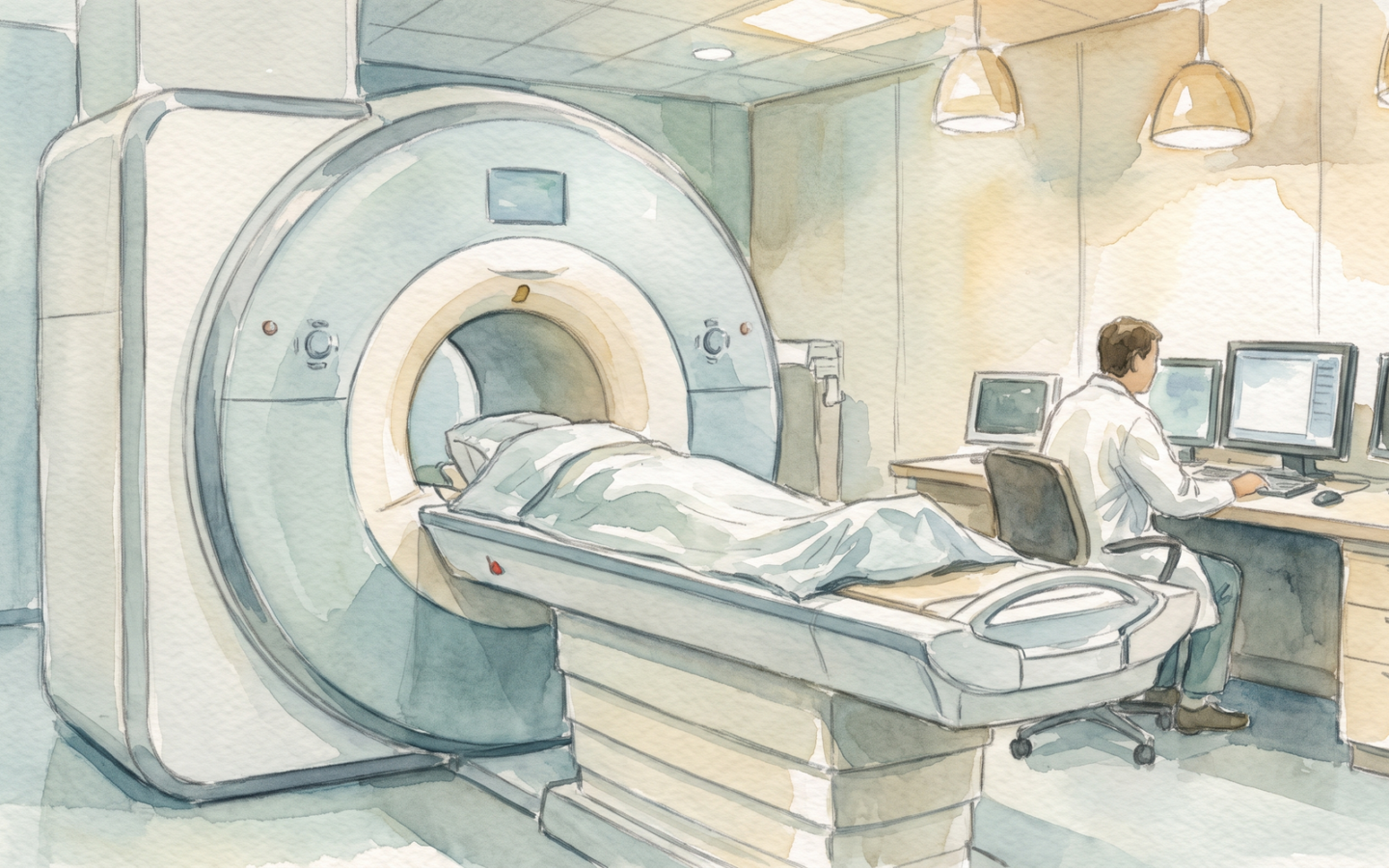
Every year, more than four billion medical imaging examinations are performed worldwide. The technologies — X-ray, CT, MRI, ultrasound, PET — have, in less than 130 years, transformed medicine from a discipline of educated guesswork to one of precise, non-invasive diagnosis. The story of how physicists, engineers, and physicians learned to look inside a living body without cutting it open is, in retrospect, one of the great triumphs of applied science.
Key findings
- X-rays were the first medical imaging modality. Wilhelm Röntgen discovered X-rays on 8 November 1895, while experimenting with a Crookes tube in his laboratory in Würzburg. Within weeks, he had taken the first medical X-ray image (of his wife’s hand, showing her wedding ring and the bones of her fingers), and within months the technology was being used in hospitals around the world. Röntgen received the first Nobel Prize in Physics in 1901 for the discovery. The basic technology — an X-ray source, an X-ray detector, and a patient between them — has changed surprisingly little in 130 years, but the sensitivity, the resolution, and the dose efficiency have improved by orders of magnitude.[1, 2, 3]
- Computed tomography (CT) made X-rays into a 3D imaging modality. The first CT scanner, built by Godfrey Hounsfield at EMI in 1971, revolutionised medical imaging. The CT scanner rotates an X-ray source and a detector around the patient, acquiring a series of 1D projections, and uses a mathematical algorithm (the Radon transform and its inverse) to reconstruct a 3D image. The first scan, of a patient’s brain, took 5 minutes to acquire and 2.5 hours to reconstruct, and produced an image with 80 × 80 pixels. The best modern CT scanners acquire a 3D volume in a fraction of a second, with spatial resolution below 0.5 mm, and with more than 1,000 slices per rotation. Hounsfield shared the 1979 Nobel Prize in Physiology or Medicine with Allan Cormack.[2, 3, 4]
- MRI uses the quantum behaviour of hydrogen nuclei to image soft tissue. Magnetic resonance imaging (MRI), independently developed by Paul Lauterbur and Peter Mansfield in the early 1970s, uses the fact that the hydrogen nucleus (a single proton) has a magnetic moment and a quantum-mechanical property called spin. When the body is placed in a strong magnetic field (typically 1.5 or 3 Tesla, more than 30,000 times the Earth’s field), the hydrogen nuclei align with the field, and can be tipped out of alignment by a radio-frequency pulse. As they re-align, they emit a radio signal that can be detected and used to form an image. The signal depends on the local chemical environment of the hydrogen, which varies between different types of tissue (fat, water, muscle, bone), giving MRI its extraordinary soft-tissue contrast. Lauterbur and Mansfield shared the 2003 Nobel Prize in Physiology or Medicine.[3, 4, 5]
- Ultrasound is the safest, cheapest, and most widely used modality. Ultrasound imaging uses high-frequency sound waves (typically 2–15 MHz, far above human hearing) that are transmitted into the body by a transducer, reflected by the internal structures, and received by the same transducer. The time of flight gives the depth of the structure, and the strength of the reflection gives the acoustic impedance (a property related to the density and the stiffness of the tissue). Ultrasound is real-time, portable, and uses no ionising radiation, making it the safest imaging modality. It is used for obstetrics, cardiology, abdominal imaging, and many other applications. The dominant ultrasound manufacturers, in 2024, are GE HealthCare, Philips, Siemens Healthineers, and Canon Medical Systems.[4, 5, 6]
- PET and SPECT image metabolism, not anatomy. Positron emission tomography (PET) and single-photon emission computed tomography (SPECT) are functional imaging modalities: they image the metabolism or the biochemistry of the tissue, rather than the anatomy. The patient is injected with a small amount of a radioactive tracer (typically a sugar analogue labelled with a positron-emitting isotope such as fluorine-18), and the scanner detects the gamma rays emitted as the tracer decays. The resulting image shows the uptake of the tracer, which is high in tissues that are metabolically active (the brain, the heart, and — unfortunately — many tumours). PET is now routinely combined with CT (PET/CT) and with MRI (PET/MRI) to provide both functional and anatomical information in a single scan.[5, 6, 7]
- AI is transforming medical imaging. The dominant trend in 2024 is the integration of deep learning into every modality. AI is now used to reconstruct CT and MRI images from less data (faster scans, lower doses), to detect and segment lesions in radiographs, CT, and MRI, to classify skin lesions in dermatology, and to triage urgent cases in emergency departments. The first AI-based medical imaging systems have been approved by regulators in the US (FDA), Europe (CE), and other jurisdictions, and the use of AI in medical imaging is growing rapidly. The long-term implications are profound: a future in which a radiologist’s work is augmented (or, in some cases, replaced) by a deep neural network, trained on millions of labelled images, is no longer science fiction.[6, 7, 8]
X-rays: the first medical imaging
The discovery of X-rays by Wilhelm Röntgen in 1895 was, in retrospect, a stroke of luck. Röntgen was experimenting with a Crookes tube — a glass tube with two electrodes and a partial vacuum, used to study cathode rays — when he noticed that a screen coated with barium platinocyanide, lying on a bench several feet away, was glowing. The Crookes tube was covered with black cardboard, so no visible light could have reached the screen. Röntgen realised that some unknown radiation, capable of passing through the cardboard, was being emitted from the tube. He called the unknown radiation X-rays, using the mathematical symbol for the unknown.[1, 2, 9]
Within weeks of the discovery, Röntgen had taken the first medical X-ray image — of his wife’s hand, showing the bones and her wedding ring — and within months the technology was being used in hospitals around the world. The medical impact was immediate and enormous: for the first time, physicians could see inside a living body without cutting it open. Broken bones, foreign objects (bullets, swallowed coins), and bone tumours could be diagnosed with certainty. The first battlefield X-ray unit was deployed in the Italo-Turkish War of 1911, and X-ray units became standard in field hospitals during the First World War. The basic technology — an X-ray source, an X-ray detector, and the patient between them — has not changed in 130 years, but the sensitivity, the resolution, and the dose efficiency have improved by orders of magnitude.[1, 2, 9]
The physics of a medical X-ray is straightforward. The X-ray source is a vacuum tube with a heated cathode (which emits electrons) and a metal anode (typically tungsten). A high voltage (30–150 kV) accelerates the electrons to a high energy, and they strike the anode, producing X-rays by bremsstrahlung (braking radiation) and by characteristic emission. The X-rays are filtered (to remove the low-energy photons, which would be absorbed in the skin and contribute nothing to the image) and collimated (to direct them at the region of interest). The X-rays pass through the body, where they are absorbed differently by different tissues: bone absorbs more than muscle, which absorbs more than fat, which absorbs more than air. The transmitted X-rays strike a detector (originally a photographic film, now a digital flat-panel detector), and the resulting image is a map of the X-ray attenuation of the body. The image is a 2D projection of a 3D object, and the depth information is lost.[1, 2, 9]
Computed tomography: X-rays in three dimensions
The computed tomography (CT) scanner was invented by Godfrey Hounsfield at EMI in 1971. The CT scanner rotates an X-ray source and a detector around the patient, acquiring a series of 1D projections at many angles. The 1D projections are combined, using a mathematical algorithm (the inverse Radon transform), to reconstruct a 2D cross-sectional image (a “slice”) of the body. By acquiring many slices in sequence, the CT scanner can produce a 3D image of the entire body. The first CT scan, of a patient’s brain, took 5 minutes to acquire and 2.5 hours to reconstruct, and produced an image with 80 × 80 pixels. The best modern CT scanners acquire a 3D volume in a fraction of a second, with spatial resolution below 0.5 mm, and with more than 1,000 slices per rotation. Hounsfield shared the 1979 Nobel Prize in Physiology or Medicine with Allan Cormack.[2, 3, 10]
The mathematics of CT reconstruction is the inverse of the Radon transform, which states that a 2D object can be uniquely reconstructed from an infinite set of 1D projections at all angles. The practical implementation uses the filtered back-projection algorithm, in which each projection is filtered with a high-pass filter (to sharpen the edges) and then smeared back across the image. The smearing of all the filtered projections, accumulated over all angles, converges to the original image. The algorithm is computationally efficient, and the modern CT scanner uses a variant called iterative reconstruction, which models the physics of the X-ray interaction and the noise of the detector to produce a higher-quality image from a lower X-ray dose.[2, 3, 10]
The clinical impact of CT has been enormous. The CT scan is now the workhorse of emergency medicine, oncology, and trauma surgery, and more than 100 million CT scans are performed worldwide each year. The downside is the radiation dose: a single CT scan of the chest delivers a radiation dose of about 7 millisieverts, comparable to the natural background radiation a person receives in two years. The dose has been falling as the technology has improved — a modern low-dose CT scan of the chest delivers less than 1 millisievert — but the cumulative dose from multiple scans is still a concern, particularly for children. The trend, in 2024, is toward low-dose CT (using AI-based reconstruction to recover image quality from a lower X-ray dose) and toward MRI and ultrasound for applications where the radiation dose is a concern.[2, 3, 10]
MRI: the quantum behaviour of hydrogen
Magnetic resonance imaging (MRI) uses the quantum behaviour of the hydrogen nucleus (a single proton) to image soft tissue. The hydrogen nucleus has a magnetic moment (it behaves like a tiny bar magnet) and a quantum-mechanical property called spin. When the body is placed in a strong magnetic field (typically 1.5 or 3 Tesla, more than 30,000 times the Earth’s magnetic field), the hydrogen nuclei align with the field, with slightly more aligned parallel than anti-parallel. The aligned nuclei can be tipped out of alignment by a radio-frequency pulse at a specific frequency (the Larmor frequency, which is proportional to the magnetic field). As the nuclei re-align, they emit a radio signal that can be detected by a receiver coil. The signal depends on the local chemical environment of the hydrogen, which varies between different types of tissue (fat, water, muscle, bone), giving MRI its extraordinary soft-tissue contrast.[3, 4, 11]
The key insight of MRI is the use of gradient magnetic fields to spatially encode the signal. By applying a gradient in the magnetic field along one direction (the so-called “slice-select” gradient), only the nuclei in a thin slice of the body resonate at the Larmor frequency, and only those nuclei contribute to the signal. By applying additional gradients during the signal acquisition (the “frequency-encoding” and “phase-encoding” gradients), the spatial position of the nuclei in the slice can be reconstructed using a Fourier transform. The result is a 2D image of a thin slice of the body, and by acquiring many slices, a 3D image. The whole acquisition is governed by a “pulse sequence”, which specifies the timing and amplitude of the radio-frequency pulses and the gradients. There are many pulse sequences, each optimised for a particular type of tissue or a particular clinical question.[3, 4, 11]
The clinical impact of MRI has been equally enormous. MRI is now the gold standard for imaging the brain, the spine, the joints, and the soft tissues of the abdomen and pelvis. More than 100 million MRI scans are performed worldwide each year. The downsides are the cost (an MRI scanner costs $1–3 million, and a scan costs $500–$3,000), the scan time (15–60 minutes per scan, vs seconds for CT), and the contraindications (patients with certain metallic implants, including some pacemakers, cannot be scanned). The trend, in 2024, is toward higher field strengths (7 Tesla scanners are now available, with even better resolution and contrast), toward faster scans (using AI-based reconstruction to recover image quality from less data), and toward portable MRI (low-field scanners that can be brought to the bedside in the ICU).[3, 4, 11]
Ultrasound: the safest, cheapest, most versatile
Ultrasound imaging uses high-frequency sound waves, typically 2–15 MHz (far above the range of human hearing, which is 20 Hz to 20 kHz), to image the inside of the body. The sound waves are generated and detected by a piezoelectric transducer, which converts electrical energy to mechanical vibrations and back. The transducer is placed on the skin, with a layer of gel to couple the sound into the body, and the sound waves propagate into the body, where they are reflected by the internal structures. The time of flight gives the depth of the structure, and the strength of the reflection gives the acoustic impedance (a property related to the density and the stiffness of the tissue). The reflected waves are detected by the same transducer, and the resulting image is a 2D map of the acoustic impedance of the body.[4, 5, 12]
The advantages of ultrasound are its safety, its cost, and its versatility. The sound waves are non-ionising (they do not damage DNA), so ultrasound is safe for use in obstetrics, paediatrics, and other applications where radiation is a concern. The transducers are cheap (a few hundred dollars for a basic probe), and the scanners are portable (a modern portable ultrasound scanner weighs less than 1 kg and costs less than $5,000). The scan is real-time (the image is updated 30–60 times per second), allowing the operator to see the motion of the heart, the flow of blood, the movement of a fetus. The downside is the limited resolution and the limited penetration: the resolution improves with frequency, but the penetration decreases with frequency, so high-frequency ultrasound (15 MHz) can resolve structures less than 0.5 mm in size, but only to a depth of a few centimetres; low-frequency ultrasound (2 MHz) can penetrate to a depth of 30 cm, but the resolution is only a few millimetres.[4, 5, 12]
The dominant ultrasound manufacturers, in 2024, are GE HealthCare, Philips, Siemens Healthineers, and Canon Medical Systems. The technology is mature, but the integration of AI is now driving a new generation of features: AI-based auto-measurement (the scanner automatically measures the size of a tumour or the ejection fraction of the heart), AI-based image quality enhancement (the scanner cleans up the image in real time), and AI-based diagnosis (the scanner highlights suspicious regions for the sonographer to focus on). The use of AI in ultrasound is, in 2024, one of the fastest-growing areas of medical imaging, and the technology is expected to become the dominant diagnostic tool in primary care and emergency medicine over the next decade.[4, 5, 12]
PET and SPECT: imaging metabolism
Positron emission tomography (PET) and single-photon emission computed tomography (SPECT) are functional imaging modalities: they image the metabolism or the biochemistry of the tissue, rather than the anatomy. The patient is injected with a small amount of a radioactive tracer, and the scanner detects the gamma rays emitted as the tracer decays. The most common PET tracer is fluorodeoxyglucose (FDG), a sugar analogue labelled with fluorine-18, which is taken up by metabolically active tissues (the brain, the heart, and — unfortunately — many tumours). The PET scanner detects the two 511 keV gamma rays emitted when the positron from the FDG decay annihilates with an electron, and uses the coincidence of the two detections to localise the decay. The result is a 3D image of the FDG uptake in the body.[5, 6, 13]
PET is now routinely combined with CT (PET/CT) and with MRI (PET/MRI) to provide both functional and anatomical information in a single scan. PET/CT is the standard of care for cancer staging, for the diagnosis of many neurological conditions (Alzheimer’s, Parkinson’s, epilepsy), and for cardiac imaging. The downside is the cost (a PET scanner costs $2–5 million, and a PET/CT scan costs $2,000–$5,000) and the radiation dose (the FDG injection delivers about 7 millisieverts, and the CT scan adds another 7–15 millisieverts). The trend, in 2024, is toward lower-dose tracers, toward longer-lived tracers (which can be shipped from a central cyclotron to hospitals without one), and toward AI-based reconstruction (which can recover image quality from a lower dose).[5, 6, 13]
SPECT is similar to PET but uses single-photon-emitting tracers (typically technetium-99m, which is cheaper and more widely available than the positron-emitting tracers used in PET). The SPECT scanner uses a gamma camera, which is cheaper than a PET scanner but has lower resolution. SPECT is widely used for cardiac imaging (myocardial perfusion imaging), for bone imaging, and for some neurological applications. The dominant SPECT radiotracer, technetium-99m sestamibi, is used to image the blood flow to the heart muscle. The trend, in 2024, is toward hybrid SPECT/CT scanners, which combine the functional information of SPECT with the anatomical information of CT.[5, 6, 13]
AI and the future of medical imaging
The dominant trend in medical imaging, in 2024, is the integration of deep learning into every modality. AI is now used to reconstruct CT and MRI images from less data (faster scans, lower doses), to detect and segment lesions in radiographs, CT, and MRI, to classify skin lesions in dermatology, and to triage urgent cases in emergency departments. The first AI-based medical imaging systems have been approved by regulators in the US (FDA), Europe (CE), and other jurisdictions, and the use of AI in medical imaging is growing rapidly.[6, 7, 14]
The most successful application, so far, is in radiology. AI systems are now used to detect diabetic retinopathy in retinal photographs, to detect breast cancer in mammograms, to detect lung cancer in low-dose CT, and to detect stroke in CT of the brain. The performance of the best AI systems, in 2024, is comparable to that of expert radiologists, and in some cases (the detection of small lesions, the integration of multiple imaging modalities) it is better. The radiologist’s role is changing, from “find the lesion” to “verify the AI’s finding, integrate it with the clinical context, and communicate it to the patient”. The transformation is, in many ways, comparable to the introduction of the PACS (Picture Archiving and Communication System) in the 1990s, which moved radiology from film to digital.[6, 7, 14]
The long-term implications are profound. A future in which every imaging examination is automatically analysed by an AI, with the results reviewed by a radiologist only when the AI is uncertain, is now technically feasible. The cost of imaging could fall dramatically, and the access to imaging could expand to underserved areas. The radiologist’s role would shift from primary diagnosis to consultation, to complex case management, and to the integration of imaging with the broader clinical context. The transformation, in 2024, is in its early stages, and the regulatory, legal, and ethical frameworks are still being developed. But the trajectory is clear: AI will, in the next decade, transform medical imaging in the same way that the introduction of X-rays did in 1895.[6, 7, 15]
Comparison: leading medical imaging modalities (2024)
| Modality | Physical principle | Best for | Typical resolution | Radiation dose | Scan time | Cost (US$) |
|---|---|---|---|---|---|---|
| X-ray (radiograph) | X-ray attenuation | Bones, lungs, foreign bodies | ~0.1 mm | ~0.1 mSv | <1 second | $50–200 |
| CT scan | X-ray attenuation, multi-angle | Trauma, chest, abdomen, oncology | ~0.5 mm | ~7 mSv (chest) | seconds | $300–3,000 |
| MRI | Nuclear magnetic resonance | Brain, spine, joints, soft tissue | ~1 mm | 0 (non-ionising) | 15–60 min | $500–3,000 |
| Ultrasound | Acoustic reflection | Obstetrics, cardiology, abdomen, vascular | ~0.5 mm (at 15 MHz) | 0 (non-ionising) | real-time | $100–500 |
| PET (with FDG) | Positron annihilation | Oncology, brain, cardiac metabolism | ~4 mm | ~7 mSv (FDG) + 7 mSv (CT) | 20–40 min | $2,000–5,000 |
| SPECT | Single-photon emission | Cardiac, bone, thyroid | ~7 mm | ~10 mSv | 15–30 min | $500–2,000 |
Key numbers
- ~4 billion: medical imaging examinations performed worldwide each year (estimated).
- ~100 million: CT scans performed worldwide each year.
- ~100 million: MRI scans performed worldwide each year.
- ~$150 billion: global medical imaging market (2024).
- ~7 mSv: radiation dose from a single chest CT scan.
- ~30,000x: ratio of MRI’s magnetic field to Earth’s.
- 1895: year of the discovery of X-rays.
- 1971: year of the first CT scan.
- 1977: year of the first whole-body MRI scan.
References
- Wikipedia, “X-ray.” (Standard reference on X-rays, with a section on the discovery.) https://en.wikipedia.org/wiki/X-ray
- Wikipedia, “Wilhelm Röntgen.” (Standard reference on Wilhelm Röntgen, the discoverer of X-rays.) https://en.wikipedia.org/wiki/Wilhelm_R%C3%B6ntgen
- Wikipedia, “CT scan.” (Standard reference on CT, with a section on the history.) https://en.wikipedia.org/wiki/CT_scan
- Wikipedia, “Magnetic resonance imaging.” (Standard reference on MRI, with a section on the physics.) https://en.wikipedia.org/wiki/Magnetic_resonance_imaging
- Wikipedia, “Ultrasound.” (Standard reference on medical ultrasound, with a section on the history.) https://en.wikipedia.org/wiki/Ultrasound
- Wikipedia, “Positron emission tomography.” (Standard reference on PET, with a section on the most common tracers.) https://en.wikipedia.org/wiki/Positron_emission_tomography
- Wikipedia, “Artificial intelligence in healthcare.” (Standard reference on the application of AI in healthcare, with a section on medical imaging.) https://en.wikipedia.org/wiki/Artificial_intelligence_in_healthcare
- Wikipedia, “Godfrey Hounsfield.” (Standard reference on Godfrey Hounsfield, the inventor of the CT scanner.) https://en.wikipedia.org/wiki/Godfrey_Hounsfield
- Wikipedia, “Bremsstrahlung.” (Standard reference on bremsstrahlung, the mechanism by which X-rays are produced in a medical X-ray tube.) https://en.wikipedia.org/wiki/Bremsstrahlung
- Wikipedia, “Radon transform.” (Standard reference on the Radon transform, the mathematical basis of CT reconstruction.) https://en.wikipedia.org/wiki/Radon_transform
- Wikipedia, “Paul Lauterbur.” (Standard reference on Paul Lauterbur, the inventor of MRI.) https://en.wikipedia.org/wiki/Paul_Lauterbur
- Wikipedia, “GE HealthCare.” (Standard reference on GE HealthCare, the leading medical imaging company.) https://en.wikipedia.org/wiki/GE_HealthCare
- Wikipedia, “Fludeoxyglucose (18F).” (Standard reference on FDG, the most common PET tracer.) https://en.wikipedia.org/wiki/Fludeoxyglucose_(18F)
- Wikipedia, “Picture archiving and communication system.” (Standard reference on PACS, the system that moved radiology from film to digital.) https://en.wikipedia.org/wiki/Picture_archiving_and_communication_system
- Wikipedia, “Diabetic retinopathy.” (Standard reference on diabetic retinopathy, the first condition for which an AI-based diagnostic system was widely deployed.) https://en.wikipedia.org/wiki/Diabetic_retinopathy